4BFI

Structure of the complex of the extracellular portions of mouse CD200R and mouse CD200


Experimental Data Snapshot

  • Method: X-RAY DIFFRACTION
  • Resolution: 3.22 Å
  • R-Value Free: 0.251 
  • R-Value Work: 0.199 
  • R-Value Observed: 0.202 

wwPDB Validation   3D Report Full Report


Ligand Structure Quality Assessment 


This is version 1.4 of the entry. See complete history


Literature

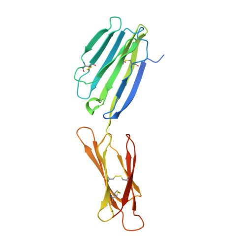
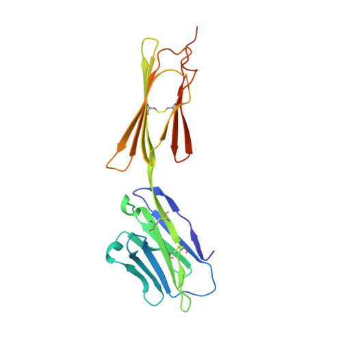
Structures of Cd200/Cd200 Receptor Family and Implications for Topology, Regulation, and Evolution

Hatherley, D.Lea, S.M.Johnson, S.Barclay, A.N.

(2013) Structure 21: 820

  • DOI: https://doi.org/10.1016/j.str.2013.03.008
  • Primary Citation of Related Structures:  
    4BFE, 4BFG, 4BFI

  • PubMed Abstract: 

    CD200 is a widely distributed membrane glycoprotein that regulates myeloid cell activity through its interaction with an inhibitory receptor (CD200R). The interaction is of interest as a target for treating excessive inflammation and for treating leukemia. There are closely related proteins to CD200R that give activating signals making this a "paired receptor." We report X-ray crystallography structures for the inhibitory CD200R, the activating receptor CD200RLa, and a complex between CD200R and CD200. Both CD200 and CD200R contain two Ig-like domains and interact through their NH₂ terminal domains compatible with immunological synapse-like interactions occurring between myeloid cells and other CD200-expressing cells. The failure of the activating receptor to bind CD200 resides in subtle changes around the interface. CD200 has been acquired by herpes viruses to mimic the host interaction. CD200R has evolved rapidly presumably driven by pathogen pressure but it may also be important in homeostasis through interactions with commensal bacteria.


  • Organizational Affiliation

    Sir William Dunn School of Pathology, University of Oxford, South Parks Road, Oxford OX1 3RE, UK.


Macromolecules
Find similar proteins by:  (by identity cutoff)  |  3D Structure
Entity ID: 1
MoleculeChains Sequence LengthOrganismDetailsImage
CELL SURFACE GLYCOPROTEIN CD200 RECEPTOR 1216Mus musculusMutation(s): 0 
UniProt
Find proteins for Q9ES57 (Mus musculus)
Explore Q9ES57 
Go to UniProtKB:  Q9ES57
Entity Groups  
Sequence Clusters30% Identity50% Identity70% Identity90% Identity95% Identity100% Identity
UniProt GroupQ9ES57
Sequence Annotations
Expand
  • Reference Sequence
Find similar proteins by:  (by identity cutoff)  |  3D Structure
Entity ID: 2
MoleculeChains Sequence LengthOrganismDetailsImage
OX-2 MEMBRANE GLYCOPROTEIN211Mus musculusMutation(s): 0 
UniProt & NIH Common Fund Data Resources
Find proteins for O54901 (Mus musculus)
Explore O54901 
Go to UniProtKB:  O54901
IMPC:  MGI:1196990
Entity Groups  
Sequence Clusters30% Identity50% Identity70% Identity90% Identity95% Identity100% Identity
UniProt GroupO54901
Sequence Annotations
Expand
  • Reference Sequence
Small Molecules
Ligands 3 Unique
IDChains Name / Formula / InChI Key2D Diagram3D Interactions
NAG
Query on NAG

Download Ideal Coordinates CCD File 
C [auth A]
D [auth A]
G [auth A]
H [auth A]
I [auth A]
C [auth A],
D [auth A],
G [auth A],
H [auth A],
I [auth A],
J [auth B],
K [auth B],
L [auth B],
M [auth B],
N [auth B],
O [auth B]
2-acetamido-2-deoxy-beta-D-glucopyranose
C8 H15 N O6
OVRNDRQMDRJTHS-FMDGEEDCSA-N
CYS
Query on CYS

Download Ideal Coordinates CCD File 
F [auth A]CYSTEINE
C3 H7 N O2 S
XUJNEKJLAYXESH-REOHCLBHSA-N
EDO
Query on EDO

Download Ideal Coordinates CCD File 
E [auth A]1,2-ETHANEDIOL
C2 H6 O2
LYCAIKOWRPUZTN-UHFFFAOYSA-N
Experimental Data & Validation

Experimental Data

  • Method: X-RAY DIFFRACTION
  • Resolution: 3.22 Å
  • R-Value Free: 0.251 
  • R-Value Work: 0.199 
  • R-Value Observed: 0.202 
  • Space Group: P 43 21 2
Unit Cell:
Length ( Å )Angle ( ˚ )
a = 128.17α = 90
b = 128.17β = 90
c = 116.08γ = 90
Software Package:
Software NamePurpose
REFMACrefinement
XDSdata reduction
XSCALEdata scaling
PHASERphasing

Structure Validation

View Full Validation Report


Ligand Structure Quality Assessment 


Entry History 

Deposition Data

Revision History  (Full details and data files)

  • Version 1.0: 2013-05-01
    Type: Initial release
  • Version 1.1: 2013-05-08
    Changes: Database references
  • Version 1.2: 2013-05-29
    Changes: Database references
  • Version 1.3: 2020-07-29
    Type: Remediation
    Reason: Carbohydrate remediation
    Changes: Data collection, Derived calculations, Other, Structure summary
  • Version 1.4: 2023-12-20
    Changes: Data collection, Database references, Refinement description, Structure summary