4BAG

Feruloyl Esterase Domain of XYNY from Clostridium thermocellum after exposure to 266nm UV laser


Experimental Data Snapshot

  • Method: X-RAY DIFFRACTION
  • Resolution: 1.90 Å
  • R-Value Free: 0.187 
  • R-Value Work: 0.167 
  • R-Value Observed: 0.168 

wwPDB Validation   3D Report Full Report


This is version 2.1 of the entry. See complete history


Literature

In-House Uv Radiation-Damage-Induced Phasing of Selenomethionine-Labeled Protein Structures.

Pereira, P.J.B.Royant, A.Panjikar, S.De Sanctis, D.

(2013) J Struct Biol 181: 89

  • DOI: https://doi.org/10.1016/j.jsb.2012.11.003
  • Primary Citation of Related Structures:  
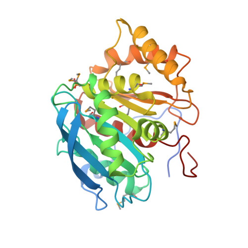
    4BAG, 4BAI, 4BAJ, 4H35

  • PubMed Abstract: 

    Selenomethionine labeling is the most common technique used in protein crystallography to derivatize recombinant proteins for experimental phasing using anomalous scattering at tunable synchrotron beamlines. Recently, it has been shown that UV radiation depletes electron density of selenium atoms of selenomethionine residues and that UV radiation-damage-induced phasing (equivalent to single isomorphous replacement) protocol can be applied to calculate experimental phases. Here we present the straightforward integration of a UV source with an in-house diffractometer. We show how this setup can extend the capabilities of a sealed tube X-ray generator and be used for experimental phasing of selenium-labeled proteins.


  • Organizational Affiliation

    IBMC-Instituto de Biologia Molecular e Celular, Universidade do Porto, Rua do Campo Alegre 823, 4150-180 Porto, Portugal.


Macromolecules
Find similar proteins by:  (by identity cutoff)  |  3D Structure
Entity ID: 1
MoleculeChains Sequence LengthOrganismDetailsImage
ENDO-1,4-BETA-XYLANASE Y
A, B
297Acetivibrio thermocellusMutation(s): 0 
EC: 3.2.1.8
UniProt
Find proteins for P51584 (Acetivibrio thermocellus)
Explore P51584 
Go to UniProtKB:  P51584
Entity Groups  
Sequence Clusters30% Identity50% Identity70% Identity90% Identity95% Identity100% Identity
UniProt GroupP51584
Sequence Annotations
Expand
  • Reference Sequence
Small Molecules
Modified Residues  2 Unique
IDChains TypeFormula2D DiagramParent
MSE
Query on MSE
A, B
L-PEPTIDE LINKINGC5 H11 N O2 SeMET
SEP
Query on SEP
A, B
L-PEPTIDE LINKINGC3 H8 N O6 PSER
Experimental Data & Validation

Experimental Data

  • Method: X-RAY DIFFRACTION
  • Resolution: 1.90 Å
  • R-Value Free: 0.187 
  • R-Value Work: 0.167 
  • R-Value Observed: 0.168 
  • Space Group: P 21 21 21
Unit Cell:
Length ( Å )Angle ( ˚ )
a = 65.37α = 90
b = 108.4β = 90
c = 112.86γ = 90
Software Package:
Software NamePurpose
PHENIXrefinement
XDSdata reduction
XSCALEdata scaling
SHELXphasing

Structure Validation

View Full Validation Report


Entry History 

Deposition Data

Revision History  (Full details and data files)

  • Version 1.0: 2013-04-17
    Type: Initial release
  • Version 2.0: 2018-04-04
    Changes: Atomic model, Data collection
  • Version 2.1: 2018-06-06
    Changes: Data collection, Refinement description