3R5K

A designed redox-controlled caspase-7


Experimental Data Snapshot

  • Method: X-RAY DIFFRACTION
  • Resolution: 2.86 Å
  • R-Value Free: 0.251 
  • R-Value Work: 0.188 
  • R-Value Observed: 0.191 

wwPDB Validation   3D Report Full Report


This is version 1.3 of the entry. See complete history


Literature

A designed redox-controlled caspase.

Witkowski, W.A.Hardy, J.A.

(2011) Protein Sci 20: 1421-1431

  • DOI: https://doi.org/10.1002/pro.673
  • Primary Citation of Related Structures:  
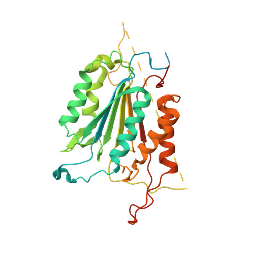
    3R5K

  • PubMed Abstract: 

    Caspases are a powerful class of cysteine proteases. Introduction of activated caspases in healthy or cancerous cells results in induction of apoptotic cell death. In this study, we have designed and characterized a version of caspase-7 that can be inactivated under oxidizing extracellular conditions and then reactivated under reducing intracellular conditions. This version of caspase-7 is allosterically inactivated when two of the substrate-binding loops are locked together via an engineered disulfide. When this disulfide is reduced, the protein regains its full function. The inactive loop-locked version of caspase-7 can be readily observed by immunoblotting and mass spectrometry. The reduced and reactivated form of the enzyme observed crystallographically is the first caspase-7 structure in which the substrate-binding groove is properly ordered even in the absence of an active-site ligand. In the reactivated structure, the catalytic-dyad cysteine-histidine are positioned 3.5 Å apart in an orientation that is capable of supporting catalysis. This redox-controlled version of caspase-7 is particularly well suited for targeted cell death in concert with redox-triggered delivery vehicles.


  • Organizational Affiliation

    Department of Chemistry, University of Massachusetts Amherst, Amherst, Massachusetts 01003, USA.


Macromolecules
Find similar proteins by:  (by identity cutoff)  |  3D Structure
Entity ID: 1
MoleculeChains Sequence LengthOrganismDetailsImage
Caspase-7
A, B
312Homo sapiensMutation(s): 2 
Gene Names: CASP7MCH3
EC: 3.4.22.60
UniProt & NIH Common Fund Data Resources
Find proteins for P55210 (Homo sapiens)
Explore P55210 
Go to UniProtKB:  P55210
PHAROS:  P55210
GTEx:  ENSG00000165806 
Entity Groups  
Sequence Clusters30% Identity50% Identity70% Identity90% Identity95% Identity100% Identity
UniProt GroupP55210
Sequence Annotations
Expand
  • Reference Sequence
Experimental Data & Validation

Experimental Data

  • Method: X-RAY DIFFRACTION
  • Resolution: 2.86 Å
  • R-Value Free: 0.251 
  • R-Value Work: 0.188 
  • R-Value Observed: 0.191 
  • Space Group: P 32 2 1
Unit Cell:
Length ( Å )Angle ( ˚ )
a = 89.85α = 90
b = 89.85β = 90
c = 185.91γ = 120
Software Package:
Software NamePurpose
ADSCdata collection
PHASERphasing
REFMACrefinement
XDSdata reduction
SCALAdata scaling

Structure Validation

View Full Validation Report


Entry History 

Deposition Data

Revision History  (Full details and data files)

  • Version 1.0: 2011-06-29
    Type: Initial release
  • Version 1.1: 2011-07-13
    Changes: Version format compliance
  • Version 1.2: 2012-11-28
    Changes: Database references
  • Version 1.3: 2023-09-13
    Changes: Data collection, Database references, Derived calculations, Refinement description