3I8E

Crystal Structure of DDB1 in Complex with the H-Box Motif of WDR42A


Experimental Data Snapshot

  • Method: X-RAY DIFFRACTION
  • Resolution: 3.40 Å
  • R-Value Free: 0.338 
  • R-Value Work: 0.260 
  • R-Value Observed: 0.264 

wwPDB Validation   3D Report Full Report


This is version 1.2 of the entry. See complete history


Literature

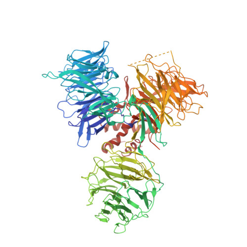
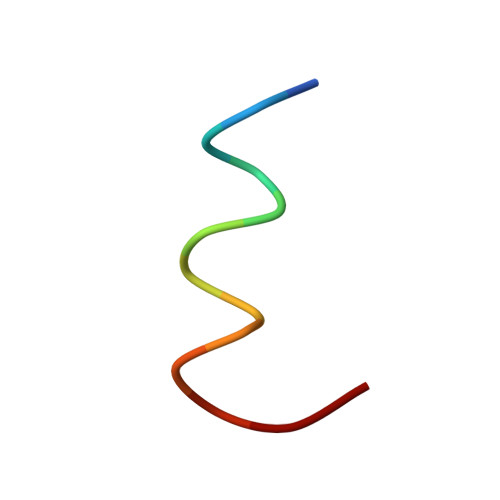
A promiscuous alpha-helical motif anchors viral hijackers and substrate receptors to the CUL4-DDB1 ubiquitin ligase machinery.

Li, T.Robert, E.I.van Breugel, P.C.Strubin, M.Zheng, N.

(2010) Nat Struct Mol Biol 17: 105-111

  • DOI: https://doi.org/10.1038/nsmb.1719
  • Primary Citation of Related Structures:  
    3I7H, 3I7K, 3I7L, 3I7N, 3I7O, 3I7P, 3I89, 3I8C, 3I8E

  • PubMed Abstract: 

    The cullin 4-DNA-damage-binding protein 1 (CUL4-DDB1) ubiquitin ligase machinery regulates diverse cellular functions and can be subverted by pathogenic viruses. Here we report the crystal structure of DDB1 in complex with a central fragment of hepatitis B virus X protein (HBx), whose DDB1-binding activity is important for viral infection. The structure reveals that HBx binds DDB1 through an alpha-helical motif, which is also found in the unrelated paramyxovirus SV5-V protein despite their sequence divergence. Our structure-based functional analysis suggests that, like SV5-V, HBx captures DDB1 to redirect the ubiquitin ligase activity of the CUL4-DDB1 E3 ligase. We also identify the alpha-helical motif shared by these viral proteins in the cellular substrate-recruiting subunits of the E3 complex, the DDB1-CUL4-associated factors (DCAFs) that are functionally mimicked by the viral hijackers. Together, our studies reveal a common yet promiscuous structural element that is important for the assembly of cellular and virally hijacked CUL4-DDB1 E3 complexes.


  • Organizational Affiliation

    Department of Pharmacology, University of Washington, Seattle, Washington, USA.


Macromolecules
Find similar proteins by:  (by identity cutoff)  |  3D Structure
Entity ID: 1
MoleculeChains Sequence LengthOrganismDetailsImage
DNA damage-binding protein 1
A, B
1,143Homo sapiensMutation(s): 0 
Gene Names: DDB1XAP1
UniProt & NIH Common Fund Data Resources
Find proteins for Q16531 (Homo sapiens)
Explore Q16531 
Go to UniProtKB:  Q16531
PHAROS:  Q16531
GTEx:  ENSG00000167986 
Entity Groups  
Sequence Clusters30% Identity50% Identity70% Identity90% Identity95% Identity100% Identity
UniProt GroupQ16531
Sequence Annotations
Expand
  • Reference Sequence

Find similar proteins by:  Sequence   |   3D Structure  

Entity ID: 2
MoleculeChains Sequence LengthOrganismDetailsImage
WD repeat-containing protein 42A
C, D
13Homo sapiensMutation(s): 0 
UniProt & NIH Common Fund Data Resources
Find proteins for Q5TAQ9 (Homo sapiens)
Explore Q5TAQ9 
Go to UniProtKB:  Q5TAQ9
PHAROS:  Q5TAQ9
GTEx:  ENSG00000132716 
Entity Groups  
Sequence Clusters30% Identity50% Identity70% Identity90% Identity95% Identity100% Identity
UniProt GroupQ5TAQ9
Sequence Annotations
Expand
  • Reference Sequence
Experimental Data & Validation

Experimental Data

  • Method: X-RAY DIFFRACTION
  • Resolution: 3.40 Å
  • R-Value Free: 0.338 
  • R-Value Work: 0.260 
  • R-Value Observed: 0.264 
  • Space Group: P 1 21 1
Unit Cell:
Length ( Å )Angle ( ˚ )
a = 64.138α = 90
b = 133.815β = 90.45
c = 183.999γ = 90
Software Package:
Software NamePurpose
HKL-2000data collection
PHASERphasing
REFMACrefinement
HKL-2000data reduction
HKL-2000data scaling

Structure Validation

View Full Validation Report


Entry History 

Deposition Data

Revision History  (Full details and data files)

  • Version 1.0: 2009-12-08
    Type: Initial release
  • Version 1.1: 2011-07-13
    Changes: Advisory, Version format compliance
  • Version 1.2: 2023-09-06
    Changes: Data collection, Database references, Refinement description