3CW2

Crystal structure of the intact archaeal translation initiation factor 2 from Sulfolobus solfataricus .


Experimental Data Snapshot

  • Method: X-RAY DIFFRACTION
  • Resolution: 2.80 Å
  • R-Value Free: 0.276 
  • R-Value Work: 0.225 

wwPDB Validation   3D Report Full Report


This is version 1.3 of the entry. See complete history


Literature

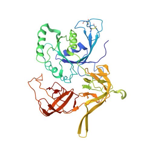
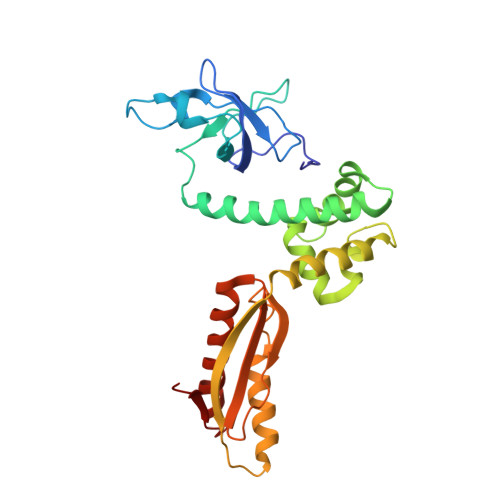
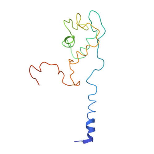
Crystal structure of the intact archaeal translation initiation factor 2 demonstrates very high conformational flexibility in the alpha- and beta-subunits.

Stolboushkina, E.Nikonov, S.Nikulin, A.Blasi, U.Manstein, D.J.Fedorov, R.Garber, M.Nikonov, O.

(2008) J Mol Biol 382: 680-691

  • DOI: https://doi.org/10.1016/j.jmb.2008.07.039
  • Primary Citation of Related Structures:  
    3CW2

  • PubMed Abstract: 

    In Eukarya and Archaea, translation initiation factor 2 (eIF2/aIF2), which contains three subunits (alpha, beta, and gamma), is pivotal for binding of charged initiator tRNA to the small ribosomal subunit. The crystal structure of the full-sized heterotrimeric aIF2 from Sulfolobus solfataricus in the nucleotide-free form has been determined at 2.8-A resolution. Superposition of four molecules in the asymmetric unit of the crystal and the comparison of the obtained structures with the known structures of the aIF2alphagamma and aIF2betagamma heterodimers revealed high conformational flexibility in the alpha- and beta-subunits. In fact, the full-sized aIF2 consists of a rigid central part, formed by the gamma-subunit, domain 3 of the alpha-subunit, and the N-terminal alpha-helix of the beta-subunit, and two mobile "wings," formed by domains 1 and 2 of the alpha-subunit, the central part, and the zinc-binding domain of the beta-subunit. High structural flexibility of the wings is probably required for interaction of aIF2 with the small ribosomal subunit. Comparative analysis of all known structures of the gamma-subunit alone and within the heterodimers and heterotrimers in nucleotide-bound and nucleotide-free states shows that the conformations of switch 1 and switch 2 do not correlate with the assembly or nucleotide states of the protein.


  • Organizational Affiliation

    Institute of Protein Research, Russian Academy of Sciences, 142290 Pushchino, Moscow Region, Russian Federation.


Macromolecules
Find similar proteins by:  (by identity cutoff)  |  3D Structure
Entity ID: 1
MoleculeChains Sequence LengthOrganismDetailsImage
Translation initiation factor 2 subunit gammaA,
B,
G [auth E],
H [auth F]
415Saccharolobus solfataricusMutation(s): 0 
Gene Names: eif2gSSO0412
UniProt
Find proteins for Q980A5 (Saccharolobus solfataricus (strain ATCC 35092 / DSM 1617 / JCM 11322 / P2))
Explore Q980A5 
Go to UniProtKB:  Q980A5
Entity Groups  
Sequence Clusters30% Identity50% Identity70% Identity90% Identity95% Identity100% Identity
UniProt GroupQ980A5
Sequence Annotations
Expand
  • Reference Sequence
Find similar proteins by:  (by identity cutoff)  |  3D Structure
Entity ID: 2
MoleculeChains Sequence LengthOrganismDetailsImage
Translation initiation factor 2 subunit alphaC,
D,
I [auth G],
J [auth H]
266Saccharolobus solfataricusMutation(s): 0 
Gene Names: eif2aSSO1050
UniProt
Find proteins for Q97Z79 (Saccharolobus solfataricus (strain ATCC 35092 / DSM 1617 / JCM 11322 / P2))
Explore Q97Z79 
Go to UniProtKB:  Q97Z79
Entity Groups  
Sequence Clusters30% Identity50% Identity70% Identity90% Identity95% Identity100% Identity
UniProt GroupQ97Z79
Sequence Annotations
Expand
  • Reference Sequence
Find similar proteins by:  (by identity cutoff)  |  3D Structure
Entity ID: 3
MoleculeChains Sequence LengthOrganismDetailsImage
Translation initiation factor 2 subunit betaE [auth K],
F [auth L],
K [auth M],
L [auth N]
139Saccharolobus solfataricusMutation(s): 0 
Gene Names: eif2bSSO2381
UniProt
Find proteins for Q97W59 (Saccharolobus solfataricus (strain ATCC 35092 / DSM 1617 / JCM 11322 / P2))
Explore Q97W59 
Go to UniProtKB:  Q97W59
Entity Groups  
Sequence Clusters30% Identity50% Identity70% Identity90% Identity95% Identity100% Identity
UniProt GroupQ97W59
Sequence Annotations
Expand
  • Reference Sequence
Experimental Data & Validation

Experimental Data

  • Method: X-RAY DIFFRACTION
  • Resolution: 2.80 Å
  • R-Value Free: 0.276 
  • R-Value Work: 0.225 
  • Space Group: P 1 21 1
Unit Cell:
Length ( Å )Angle ( ˚ )
a = 79.2α = 90
b = 162.92β = 90
c = 161.28γ = 90
Software Package:
Software NamePurpose
XDSdata scaling
PHASERphasing
PHENIXrefinement
XDSdata reduction

Structure Validation

View Full Validation Report


Entry History 

Deposition Data

Revision History  (Full details and data files)

  • Version 1.0: 2009-01-06
    Type: Initial release
  • Version 1.1: 2011-07-13
    Changes: Version format compliance
  • Version 1.2: 2018-01-24
    Changes: Structure summary
  • Version 1.3: 2023-08-30
    Changes: Data collection, Database references, Refinement description