3BOV

Crystal structure of the receptor binding domain of mouse PD-L2


Experimental Data Snapshot

  • Method: X-RAY DIFFRACTION
  • Resolution: 1.77 Å
  • R-Value Free: 0.223 
  • R-Value Work: 0.190 
  • R-Value Observed: 0.192 

wwPDB Validation   3D Report Full Report


This is version 1.3 of the entry. See complete history


Literature

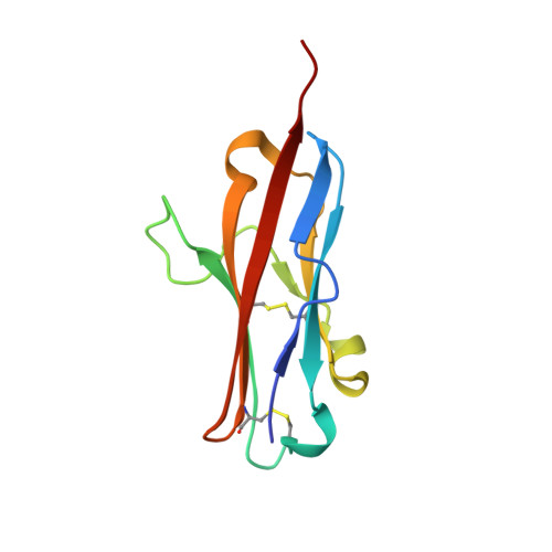
Crystal structure of the complex between programmed death-1 (PD-1) and its ligand PD-L2.

Lazar-Molnar, E.Yan, Q.Cao, E.Ramagopal, U.Nathenson, S.G.Almo, S.C.

(2008) Proc Natl Acad Sci U S A 105: 10483-10488

  • DOI: https://doi.org/10.1073/pnas.0804453105
  • Primary Citation of Related Structures:  
    3BOV, 3BP5

  • PubMed Abstract: 

    Programmed death-1 (PD-1) is a member of the CD28/B7 superfamily that delivers negative signals upon interaction with its two ligands, PD-L1 or PD-L2. The high-resolution crystal structure of the complex formed by the complete ectodomains of murine PD-1 and PD-L2 revealed a 1:1 receptor:ligand stoichiometry and displayed a binding interface and overall molecular organization distinct from that observed in the CTLA-4/B7 inhibitory complexes. Furthermore, our structure also provides insights into the association between PD-1 and PD-L1 and highlights differences in the interfaces formed by the two PD-1 ligands (PD-Ls) Mutagenesis studies confirmed the details of the proposed PD-1/PD-L binding interfaces and allowed for the design of a mutant PD-1 receptor with enhanced affinity. These studies define spatial and organizational constraints that control the localization and signaling of PD-1/PD-L complexes within the immunological synapse and provide a basis for manipulating the PD-1 pathways for immunotherapy.


  • Organizational Affiliation

    Department of Microbiology and Immunology, Albert Einstein College of Medicine, 1300 Morris Park Avenue, Bronx, NY 10461, USA.


Macromolecules
Find similar proteins by:  (by identity cutoff)  |  3D Structure
Entity ID: 1
MoleculeChains Sequence LengthOrganismDetailsImage
Programmed cell death 1 ligand 2105Mus musculusMutation(s): 0 
Gene Names: Pdcd1lg2B7dcBtdcCd273Pdl2
UniProt
Find proteins for Q9WUL5 (Mus musculus)
Explore Q9WUL5 
Go to UniProtKB:  Q9WUL5
Entity Groups  
Sequence Clusters30% Identity50% Identity70% Identity90% Identity95% Identity100% Identity
UniProt GroupQ9WUL5
Sequence Annotations
Expand
  • Reference Sequence
Experimental Data & Validation

Experimental Data

  • Method: X-RAY DIFFRACTION
  • Resolution: 1.77 Å
  • R-Value Free: 0.223 
  • R-Value Work: 0.190 
  • R-Value Observed: 0.192 
  • Space Group: C 2 2 2
Unit Cell:
Length ( Å )Angle ( ˚ )
a = 45.173α = 90
b = 116.637β = 90
c = 45.108γ = 90
Software Package:
Software NamePurpose
DENZOdata reduction
SCALEPACKdata scaling
REFMACrefinement
PDB_EXTRACTdata extraction
CBASSdata collection
HKL-2000data reduction
MOLREPphasing

Structure Validation

View Full Validation Report


Entry History 

Deposition Data

Revision History  (Full details and data files)

  • Version 1.0: 2008-07-15
    Type: Initial release
  • Version 1.1: 2011-07-13
    Changes: Version format compliance
  • Version 1.2: 2017-10-25
    Changes: Refinement description
  • Version 1.3: 2024-04-03
    Changes: Data collection, Database references, Derived calculations, Refinement description