3VNV

Complex structure of viral RNA polymerase II


Experimental Data Snapshot

  • Method: X-RAY DIFFRACTION
  • Resolution: 2.60 Å
  • R-Value Free: 0.279 
  • R-Value Work: 0.220 
  • R-Value Observed: 0.223 

wwPDB Validation   3D Report Full Report


Ligand Structure Quality Assessment 


This is version 1.3 of the entry. See complete history


Literature

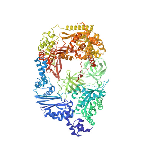
Mechanism for template-independent terminal adenylation activity of Q beta replicase

Takeshita, D.Yamashita, S.Tomita, K.

(2012) Structure 20: 1661-1669

  • DOI: https://doi.org/10.1016/j.str.2012.07.004
  • Primary Citation of Related Structures:  
    3VNU, 3VNV, 4FWT

  • PubMed Abstract: 

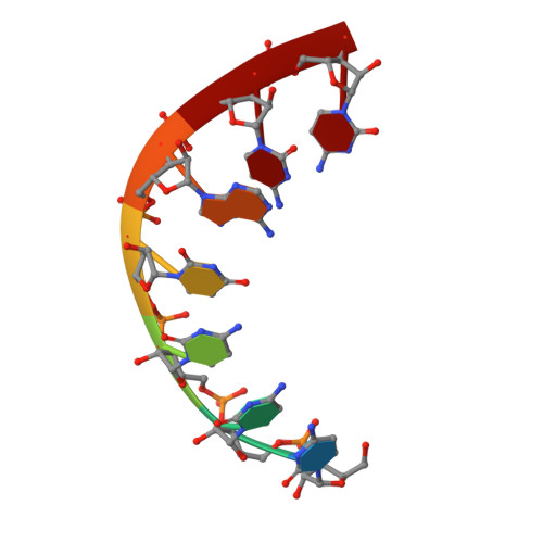
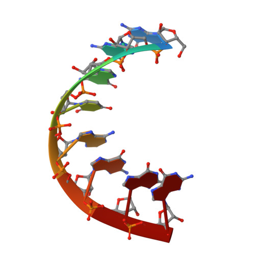
    The genomic RNA of Qβ virus is replicated by Qβ replicase, a template-dependent RNA polymerase complex. Qβ replicase has an intrinsic template-independent RNA 3'-adenylation activity, which is required for efficient viral RNA amplification in the host cells. However, the mechanism of the template-independent 3'-adenylation of RNAs by Qβ replicase has remained elusive. We determined the structure of a complex that includes Qβ replicase, a template RNA, a growing RNA complementary to the template RNA, and ATP. The structure represents the terminal stage of RNA polymerization and reveals that the shape and size of the nucleotide-binding pocket becomes available for ATP accommodation after the 3'-penultimate template-dependent C-addition. The stacking interaction between the ATP and the neighboring Watson-Crick base pair, between the 5'-G in the template and the 3'-C in the growing RNA, contributes to the nucleotide specificity. Thus, the template for the template-independent 3'-adenylation by Qβ replicase is the RNA and protein ribonucleoprotein complex.


  • Organizational Affiliation

    Biomedical Research Institute, National Institute of Advanced Industrial Science and Technology, 1-1-1, Higashi, Tsukuba, Ibaraki 305-8566, Japan.


Macromolecules

Find similar proteins by:  (by identity cutoff)  |  3D Structure
Entity ID: 1
MoleculeChains Sequence LengthOrganismDetailsImage
Elongation factor Ts, Elongation factor Tu, LINKER, Q beta replicase1,289Escherichia coli O157:H7Qubevirus durumMutation(s): 0 
UniProt
Find proteins for P14647 (Escherichia virus Qbeta)
Explore P14647 
Go to UniProtKB:  P14647
Find proteins for P0A6N3 (Escherichia coli O157:H7)
Explore P0A6N3 
Go to UniProtKB:  P0A6N3
Find proteins for P0A6P3 (Escherichia coli O157:H7)
Explore P0A6P3 
Go to UniProtKB:  P0A6P3
Entity Groups  
Sequence Clusters30% Identity50% Identity70% Identity90% Identity95% Identity100% Identity
UniProt GroupsP14647P0A6P3P0A6N3
Sequence Annotations
Expand
  • Reference Sequence

Find similar nucleic acids by:  Sequence   |   3D Structure  

Entity ID: 2
MoleculeChains LengthOrganismImage
RNA (5'-R(*CP*CP*CP*UP*AP*CP*C)-3')B [auth G]7N/A
Sequence Annotations
Expand
  • Reference Sequence

Find similar nucleic acids by:  Sequence   |   3D Structure  

Entity ID: 3
MoleculeChains LengthOrganismImage
RNA (5'-R(*GP*GP*GP*UP*AP*GP*GP*G)-3')C [auth T]8N/A
Sequence Annotations
Expand
  • Reference Sequence
Experimental Data & Validation

Experimental Data

  • Method: X-RAY DIFFRACTION
  • Resolution: 2.60 Å
  • R-Value Free: 0.279 
  • R-Value Work: 0.220 
  • R-Value Observed: 0.223 
  • Space Group: C 2 2 21
Unit Cell:
Length ( Å )Angle ( ˚ )
a = 139.73α = 90
b = 256.04β = 90
c = 101.72γ = 90
Software Package:
Software NamePurpose
ADSCdata collection
MOLREPphasing
PHENIXrefinement
HKL-2000data reduction
HKL-2000data scaling

Structure Validation

View Full Validation Report


Ligand Structure Quality Assessment 


Entry History 

Deposition Data

Revision History  (Full details and data files)

  • Version 1.0: 2012-08-08
    Type: Initial release
  • Version 1.1: 2013-10-09
    Changes: Database references
  • Version 1.2: 2017-06-28
    Changes: Source and taxonomy
  • Version 1.3: 2023-11-08
    Changes: Advisory, Data collection, Database references, Derived calculations, Refinement description