3VBA

Crystal structure of methanogen 3-isopropylmalate isomerase small subunit


Experimental Data Snapshot

  • Method: X-RAY DIFFRACTION
  • Resolution: 2.00 Å
  • R-Value Free: 0.244 
  • R-Value Work: 0.197 
  • R-Value Observed: 0.199 

wwPDB Validation   3D Report Full Report


This is version 1.1 of the entry. See complete history


Literature

Crystal structure of LeuD from Methanococcus jannaschii.

Lee, E.H.Cho, Y.W.Hwang, K.Y.

(2012) Biochem Biophys Res Commun 419: 160-164

  • DOI: https://doi.org/10.1016/j.bbrc.2012.01.125
  • Primary Citation of Related Structures:  
    3VBA

  • PubMed Abstract: 

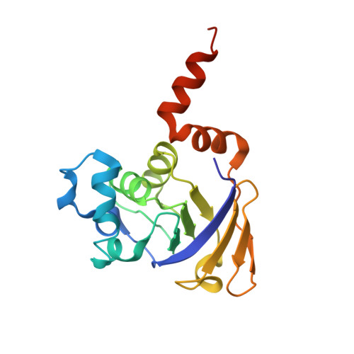
    3-Isopropylmalate/citramalate (IPM) isomerase catalyzes the second step in the leucine biosynthesis pathway. IPM isomerase from Methanococcus jannaschii is a complex protein consisting of a large (MjLeuC) and a small subunit (MjLeuD). It has broad substrate specificity, unlike other bacterial IPM isomerases. In order to understand the reasons for this broad substrate specificity, we determined the crystal structure of MjLeuD at a resolution of 2.0 Å. The asymmetric unit contained 6 molecules of LeuD, including three homodimers. The overall structure had a β/β/α sandwich-fold consisting of 8 α-helices and 7 β-strands. The C-terminal helix, which is important in homodimer formation, showed conformational differences between two homodimer forms of MjLeuD. In addition, we identified a hydrophobic residue (Val28) near the substrate recognition region that may explain the broad substrate specificity of IPM isomerase. Therefore, we suggest that LeuD proteins can be divided into 2 subfamilies, LeuD subfamilies 1 and 2, which show differences in overall structure and in the substrate recognition region.


  • Organizational Affiliation

    Division of Biotechnology, College of Life Sciences and Biotechnology, Korea University, Seoul 136-701, South Korea.


Macromolecules
Find similar proteins by:  (by identity cutoff)  |  3D Structure
Entity ID: 1
MoleculeChains Sequence LengthOrganismDetailsImage
Isopropylmalate/citramalate isomerase small subunit
A, B, C, D, E
A, B, C, D, E, F
176Methanocaldococcus jannaschii DSM 2661Mutation(s): 0 
Gene Names: leuDMJ1277
EC: 4.2.1.33 (PDB Primary Data), 4.2.1.35 (PDB Primary Data), 4.2.1.31 (PDB Primary Data)
UniProt
Find proteins for Q58673 (Methanocaldococcus jannaschii (strain ATCC 43067 / DSM 2661 / JAL-1 / JCM 10045 / NBRC 100440))
Explore Q58673 
Go to UniProtKB:  Q58673
Entity Groups  
Sequence Clusters30% Identity50% Identity70% Identity90% Identity95% Identity100% Identity
UniProt GroupQ58673
Sequence Annotations
Expand
  • Reference Sequence
Experimental Data & Validation

Experimental Data

  • Method: X-RAY DIFFRACTION
  • Resolution: 2.00 Å
  • R-Value Free: 0.244 
  • R-Value Work: 0.197 
  • R-Value Observed: 0.199 
  • Space Group: P 21 21 21
Unit Cell:
Length ( Å )Angle ( ˚ )
a = 33.836α = 90
b = 107.138β = 90
c = 288.714γ = 90
Software Package:
Software NamePurpose
PHENIXrefinement
PDB_EXTRACTdata extraction
HKL-2000data collection
HKL-2000data reduction
HKL-2000data scaling
PHENIXphasing

Structure Validation

View Full Validation Report


Entry History 

Deposition Data

Revision History  (Full details and data files)

  • Version 1.0: 2012-11-14
    Type: Initial release
  • Version 1.1: 2024-03-20
    Changes: Data collection, Database references