Structural Analysis of a Protective Epitope of the Francisella tularensis O-Polysaccharide.
Rynkiewicz, M.J., Lu, Z., Hui, J.H., Sharon, J., Seaton, B.A.(2012) Biochemistry 51: 5684-5694
- PubMed: 22747335
- DOI: https://doi.org/10.1021/bi201711m
- Primary Citation of Related Structures:
3UJT - PubMed Abstract:
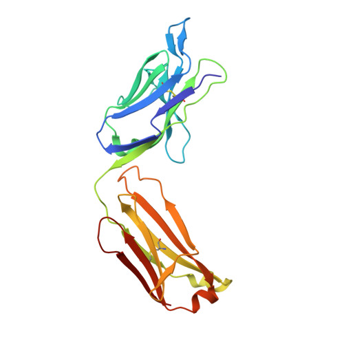
Francisella tularensis (Ft), the Gram-negative facultative intracellular bacterium that causes tularemia, is considered a biothreat because of its high infectivity and the high mortality rate of respiratory disease. The Ft lipopolysaccharide (Ft LPS) is thought to be a main protective antigen in mice and humans, and we have previously demonstrated the protective effect of the Ft LPS-specific monoclonal antibody Ab52 in a mouse model of respiratory tularemia. Immunochemical characterization has shown that the epitope recognized by Ab52 is contained within two internal repeat units of the O-polysaccharide [O-antigen (OAg)] of Ft LPS. To further localize the Ab52 epitope and understand the molecular interactions between the antibody and the saccharide, we determined the X-ray crystal structure of the Fab fragment of Ab52 and derived an antibody-antigen complex using molecular docking. The docked complex, refined through energy minimization, reveals an antigen binding site in the shape of a large canyon with a central pocket that accommodates a V-shaped epitope consisting of six sugar residues, α-D-GalpNAcAN(1→4)-α-D-GalpNAcAN(1→3)-β-D-QuipNAc(1→2)-β-D-Quip4NFm(1→4)-α-D-GalpNAcAN(1→4)-α-D-GalpNAcAN. These results inform the development of vaccines and immunotherapeutic/immunoprophylactic antibodies against Ft by suggesting a desired topology for binding of the antibody to internal epitopes of Ft LPS. This is the first report of an X-ray crystal structure of a monoclonal antibody that targets a protective Ft B cell epitope.
Organizational Affiliation:
Department of Physiology and Biophysics, Boston University School of Medicine, Boston, MA 02118, USA.