3RN6

Crystal structure of Cytosine Deaminase from Escherichia Coli complexed with zinc and isoguanine


Experimental Data Snapshot

  • Method: X-RAY DIFFRACTION
  • Resolution: 2.25 Å
  • R-Value Free: 0.243 
  • R-Value Work: 0.207 
  • R-Value Observed: 0.208 

wwPDB Validation   3D Report Full Report


Ligand Structure Quality Assessment 


This is version 1.1 of the entry. See complete history


Literature

Rescue of the orphan enzyme isoguanine deaminase.

Hitchcock, D.S.Fedorov, A.A.Fedorov, E.V.Dangott, L.J.Almo, S.C.Raushel, F.M.

(2011) Biochemistry 50: 5555-5557

  • DOI: https://doi.org/10.1021/bi200680y
  • Primary Citation of Related Structures:  
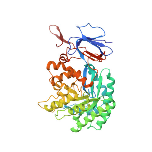
    3RN6

  • PubMed Abstract: 

    Cytosine deaminase (CDA) from Escherichia coli was shown to catalyze the deamination of isoguanine (2-oxoadenine) to xanthine. Isoguanine is an oxidation product of adenine in DNA that is mutagenic to the cell. The isoguanine deaminase activity in E. coli was partially purified by ammonium sulfate fractionation, gel filtration, and anion exchange chromatography. The active protein was identified by peptide mass fingerprint analysis as cytosine deaminase. The kinetic constants for the deamination of isoguanine at pH 7.7 are as follows: k(cat) = 49 s(-1), K(m) = 72 μM, and k(cat)/K(m) = 6.7 × 10(5) M(-1) s(-1). The kinetic constants for the deamination of cytosine are as follows: k(cat) = 45 s(-1), K(m) = 302 μM, and k(cat)/K(m) = 1.5 × 10(5) M(-1) s(-1). Under these reaction conditions, isoguanine is the better substrate for cytosine deaminase. The three-dimensional structure of CDA was determined with isoguanine in the active site.


  • Organizational Affiliation

    Department of Biochemistry and Biophysics and Department of Chemistry, Texas A&M University, College Station, Texas 77843, USA.


Macromolecules
Find similar proteins by:  (by identity cutoff)  |  3D Structure
Entity ID: 1
MoleculeChains Sequence LengthOrganismDetailsImage
Cytosine deaminase427Escherichia coliMutation(s): 0 
Gene Names: codAb0337JW0328
EC: 3.5.4.1
UniProt
Find proteins for P25524 (Escherichia coli (strain K12))
Explore P25524 
Go to UniProtKB:  P25524
Entity Groups  
Sequence Clusters30% Identity50% Identity70% Identity90% Identity95% Identity100% Identity
UniProt GroupP25524
Sequence Annotations
Expand
  • Reference Sequence
Small Molecules
Experimental Data & Validation

Experimental Data

  • Method: X-RAY DIFFRACTION
  • Resolution: 2.25 Å
  • R-Value Free: 0.243 
  • R-Value Work: 0.207 
  • R-Value Observed: 0.208 
  • Space Group: H 3 2
Unit Cell:
Length ( Å )Angle ( ˚ )
a = 147.181α = 90
b = 147.181β = 90
c = 199.747γ = 120
Software Package:
Software NamePurpose
ADSCdata collection
BALBESphasing
PHENIXrefinement
DENZOdata reduction
SCALEPACKdata scaling

Structure Validation

View Full Validation Report


Ligand Structure Quality Assessment 


Entry History 

Deposition Data

Revision History  (Full details and data files)

  • Version 1.0: 2011-08-24
    Type: Initial release
  • Version 1.1: 2023-09-13
    Changes: Data collection, Database references, Derived calculations, Refinement description