3RG2

Structure of a two-domain NRPS fusion protein containing the EntE adenylation domain and EntB aryl-carrier protein from enterobactin biosynthesis


Experimental Data Snapshot

  • Method: X-RAY DIFFRACTION
  • Resolution: 3.10 Å
  • R-Value Free: 0.264 
  • R-Value Work: 0.218 
  • R-Value Observed: 0.220 

wwPDB Validation   3D Report Full Report


Ligand Structure Quality Assessment 


This is version 1.5 of the entry. See complete history


Literature

Structural and Functional Investigation of the Intermolecular Interaction between NRPS Adenylation and Carrier Protein Domains.

Sundlov, J.A.Shi, C.Wilson, D.J.Aldrich, C.C.Gulick, A.M.

(2012) Chem Biol 19: 188-198

  • DOI: https://doi.org/10.1016/j.chembiol.2011.11.013
  • Primary Citation of Related Structures:  
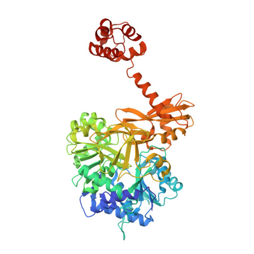
    3RG2

  • PubMed Abstract: 

    Nonribosomal peptide synthetases (NRPSs) are modular proteins that produce peptide antibiotics and siderophores. These enzymes act as catalytic assembly lines where substrates, covalently bound to integrated carrier domains, are delivered to adjacent catalytic domains. The carrier domains are initially loaded by adenylation domains, which use two distinct conformations to catalyze sequentially the adenylation of the substrate and the thioesterification of the pantetheine cofactor. We have used a mechanism-based inhibitor to determine the crystal structure of an engineered adenylation-carrier domain protein illustrating the intermolecular interaction between the adenylation and carrier domains. This structure enabled directed mutations to improve the interaction between nonnative partner proteins. Comparison with prior NRPS adenylation domain structures provides insights into the assembly line dynamics of these modular enzymes.


  • Organizational Affiliation

    Hauptman-Woodward Institute and Department of Structural Biology, University at Buffalo, Buffalo, NY 14203 USA.


Macromolecules
Find similar proteins by:  (by identity cutoff)  |  3D Structure
Entity ID: 1
MoleculeChains Sequence LengthOrganismDetailsImage
Enterobactin synthase component E (entE), 2,3-dihydro-2,3-dihydroxybenzoate synthetase, isochroismatase (Entb)
A, B, C, D, E
A, B, C, D, E, F, G, H, I, J
617Escherichia coliMutation(s): 0 
Gene Names: b0594entEentE and entBJW0586
EC: 2.7.7.58 (PDB Primary Data), 2.3.1 (PDB Primary Data)
UniProt
Find proteins for P0ADI4 (Escherichia coli (strain K12))
Explore P0ADI4 
Go to UniProtKB:  P0ADI4
Find proteins for P10378 (Escherichia coli (strain K12))
Explore P10378 
Go to UniProtKB:  P10378
Entity Groups  
Sequence Clusters30% Identity50% Identity70% Identity90% Identity95% Identity100% Identity
UniProt GroupsP0ADI4P10378
Sequence Annotations
Expand
  • Reference Sequence
Small Molecules
Ligands 3 Unique
IDChains Name / Formula / InChI Key2D Diagram3D Interactions
SVS
Query on SVS

Download Ideal Coordinates CCD File 
AA [auth D]
GA [auth E]
K [auth A]
LA [auth F]
OA [auth G]
AA [auth D],
GA [auth E],
K [auth A],
LA [auth F],
OA [auth G],
Q [auth B],
RA [auth H],
UA [auth I],
X [auth C],
ZA [auth J]
5'-deoxy-5'-({[2-(2-hydroxyphenyl)ethyl]sulfonyl}amino)adenosine
C18 H22 N6 O6 S
NIEGSPNRJDKALY-SCFUHWHPSA-N
PNS
Query on PNS

Download Ideal Coordinates CCD File 
AB [auth J]
BA [auth D]
HA [auth E]
L [auth A]
MA [auth F]
AB [auth J],
BA [auth D],
HA [auth E],
L [auth A],
MA [auth F],
PA [auth G],
R [auth B],
SA [auth H],
VA [auth I],
Y [auth C]
4'-PHOSPHOPANTETHEINE
C11 H23 N2 O7 P S
JDMUPRLRUUMCTL-VIFPVBQESA-N
IOD
Query on IOD

Download Ideal Coordinates CCD File 
BB [auth J]
CA [auth D]
CB [auth J]
DA [auth D]
EA [auth D]
BB [auth J],
CA [auth D],
CB [auth J],
DA [auth D],
EA [auth D],
FA [auth D],
IA [auth E],
JA [auth E],
KA [auth E],
M [auth A],
N [auth A],
NA [auth F],
O [auth A],
P [auth A],
QA [auth G],
S [auth B],
T [auth B],
TA [auth H],
U [auth B],
V [auth B],
W [auth B],
WA [auth I],
XA [auth I],
YA [auth I],
Z [auth C]
IODIDE ION
I
XMBWDFGMSWQBCA-UHFFFAOYSA-M
Binding Affinity Annotations 
IDSourceBinding Affinity
SVS PDBBind:  3RG2 Kd: 6.30e+4 (nM) from 1 assay(s)
Experimental Data & Validation

Experimental Data

  • Method: X-RAY DIFFRACTION
  • Resolution: 3.10 Å
  • R-Value Free: 0.264 
  • R-Value Work: 0.218 
  • R-Value Observed: 0.220 
  • Space Group: P 1 21 1
Unit Cell:
Length ( Å )Angle ( ˚ )
a = 160.575α = 90
b = 101.771β = 107.06
c = 240.678γ = 90
Software Package:
Software NamePurpose
PHENIXrefinement
PHASERphasing
BALBESphasing
REFMACrefinement
HKL-2000data collection
HKL-2000data reduction
HKL-2000data scaling

Structure Validation

View Full Validation Report


Ligand Structure Quality Assessment 


Entry History 

Deposition Data

Revision History  (Full details and data files)

  • Version 1.0: 2012-03-07
    Type: Initial release
  • Version 1.1: 2012-03-28
    Changes: Database references
  • Version 1.2: 2017-08-09
    Changes: Refinement description, Source and taxonomy
  • Version 1.3: 2019-07-17
    Changes: Data collection, Derived calculations, Refinement description
  • Version 1.4: 2019-08-14
    Changes: Data collection
  • Version 1.5: 2023-09-13
    Changes: Data collection, Database references, Derived calculations, Refinement description