3RC1

Crystal Structure of KijD10, a 3-ketoreductase from Actinomadura kijaniata incomplex with NADP and TDP-benzene


Experimental Data Snapshot

  • Method: X-RAY DIFFRACTION
  • Resolution: 1.71 Å
  • R-Value Free: 0.234 
  • R-Value Work: 0.204 
  • R-Value Observed: 0.205 

wwPDB Validation   3D Report Full Report


Ligand Structure Quality Assessment 


This is version 1.2 of the entry. See complete history


Literature

Combined Structural and Functional Investigation of a C-3''-Ketoreductase Involved in the Biosynthesis of dTDP-l-Digitoxose.

Kubiak, R.L.Holden, H.M.

(2011) Biochemistry 50: 5905-5917

  • DOI: https://doi.org/10.1021/bi200514b
  • Primary Citation of Related Structures:  
    3RBV, 3RC1, 3RC2, 3RC7, 3RC9, 3RCB

  • PubMed Abstract: 

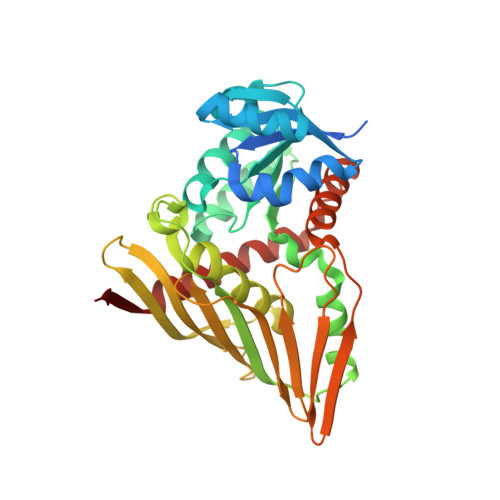
    l-Digitoxose is an unusual dideoxysugar found attached to various pharmacologically active natural products, including the antitumor antibiotic tetrocarcin A and the antibiotics kijanimicin and jadomycin B. Six enzymes are required for its production starting from glucose 1-phosphate. Here we describe a combined structural and functional investigation of KijD10, an NADPH-dependent C-3''-ketoreductase that catalyzes the third step of l-digitoxose biosynthesis in the African soil-dwelling bacterium Actinomadura kijaniata. KijD10 belongs to the glucose-fructose oxidoreductase superfamily. For this investigation, both binary and ternary complexes of KijD10 were crystallized, and their structures were determined to 2.0 Å resolution or better. On the basis of these high-resolution structures, two potential active site acids were identified, Lys 102 and Tyr 186. These residues were individually mutated and the resultant proteins investigated both kinetically and structurally. The Y186F mutant protein demonstrated significant catalytic activity, and its structure was virtually identical to that of the wild-type enzyme except for the positioning of the nicotinamide ring. All lysine mutations, on the other hand, resulted in proteins with either abolished or drastically reduced catalytic activities. Structures for the K102A and K102E mutant proteins were determined and showed that the abrogation of catalytic activity was not a result of large conformational changes. Taken together, these data suggest that Lys 102 donates a proton to the C-3'' keto group during the reaction and that Tyr 186 serves only an auxiliary role. This is in contrast to that proposed for glucose-fructose oxidoreductase and other family members in which the tyrosines, or in some cases similarly positioned histidines, are thought to play major catalytic roles.


  • Organizational Affiliation

    Department of Biochemistry, University of Wisconsin, Madison, Wisconsin 53706, United States.


Macromolecules
Find similar proteins by:  (by identity cutoff)  |  3D Structure
Entity ID: 1
MoleculeChains Sequence LengthOrganismDetailsImage
Sugar 3-ketoreductase350Actinomadura kijaniataMutation(s): 0 
Gene Names: KijD10
UniProt
Find proteins for B3TMR8 (Actinomadura kijaniata)
Explore B3TMR8 
Go to UniProtKB:  B3TMR8
Entity Groups  
Sequence Clusters30% Identity50% Identity70% Identity90% Identity95% Identity100% Identity
UniProt GroupB3TMR8
Sequence Annotations
Expand
  • Reference Sequence
Small Molecules
Ligands 6 Unique
IDChains Name / Formula / InChI Key2D Diagram3D Interactions
NAP
Query on NAP

Download Ideal Coordinates CCD File 
D [auth A]NADP NICOTINAMIDE-ADENINE-DINUCLEOTIDE PHOSPHATE
C21 H28 N7 O17 P3
XJLXINKUBYWONI-NNYOXOHSSA-N
TLO
Query on TLO

Download Ideal Coordinates CCD File 
C [auth A]5'-O-[(S)-hydroxy{[(S)-hydroxy(phenoxy)phosphoryl]oxy}phosphoryl]thymidine
C16 H20 N2 O11 P2
BANJVYKYIQPYKP-BFHYXJOUSA-N
PO4
Query on PO4

Download Ideal Coordinates CCD File 
B [auth A]PHOSPHATE ION
O4 P
NBIIXXVUZAFLBC-UHFFFAOYSA-K
EDO
Query on EDO

Download Ideal Coordinates CCD File 
F [auth A]1,2-ETHANEDIOL
C2 H6 O2
LYCAIKOWRPUZTN-UHFFFAOYSA-N
CL
Query on CL

Download Ideal Coordinates CCD File 
G [auth A],
H [auth A]
CHLORIDE ION
Cl
VEXZGXHMUGYJMC-UHFFFAOYSA-M
NA
Query on NA

Download Ideal Coordinates CCD File 
E [auth A]SODIUM ION
Na
FKNQFGJONOIPTF-UHFFFAOYSA-N
Experimental Data & Validation

Experimental Data

  • Method: X-RAY DIFFRACTION
  • Resolution: 1.71 Å
  • R-Value Free: 0.234 
  • R-Value Work: 0.204 
  • R-Value Observed: 0.205 
  • Space Group: I 2 2 2
Unit Cell:
Length ( Å )Angle ( ˚ )
a = 72.826α = 90
b = 104.154β = 90
c = 145.283γ = 90
Software Package:
Software NamePurpose
PROTEUM PLUSdata collection
PHASERphasing
REFMACrefinement
SAINTdata reduction
SADABSdata scaling

Structure Validation

View Full Validation Report


Ligand Structure Quality Assessment 


Entry History 

Deposition Data

Revision History  (Full details and data files)

  • Version 1.0: 2011-06-08
    Type: Initial release
  • Version 1.1: 2011-07-13
    Changes: Version format compliance
  • Version 1.2: 2023-09-13
    Changes: Data collection, Database references, Derived calculations, Refinement description