


Crystal structure of the complex of peptidoglycan recognition protein (PGRP-S) with dipeptide L-ALA D-GLU at 2.7 A resolution
Shukla, P.K., Sharma, P., Sinha, M., Kaur, P., Sharma, S., Singh, T.P.To be published.
Experimental Data Snapshot
Entity ID: 1 | |||||
|---|---|---|---|---|---|
| Molecule | Chains | Sequence Length | Organism | Details | Image |
| Peptidoglycan recognition protein 1 | 171 | Camelus dromedarius | Mutation(s): 0 | 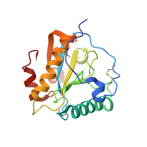 | |
UniProt | |||||
Find proteins for Q9GK12 (Camelus dromedarius) Explore Q9GK12 Go to UniProtKB: Q9GK12 | |||||
Entity Groups | |||||
| Sequence Clusters | 30% Identity50% Identity70% Identity90% Identity95% Identity100% Identity | ||||
| UniProt Group | Q9GK12 | ||||
Sequence AnnotationsExpand | |||||
| |||||
| Ligands 6 Unique | |||||
|---|---|---|---|---|---|
| ID | Chains | Name / Formula / InChI Key | 2D Diagram | 3D Interactions | |
| SRT Query on SRT | N [auth D] | S,R MESO-TARTARIC ACID C4 H6 O6 FEWJPZIEWOKRBE-XIXRPRMCSA-N |  | ||
| DGL Query on DGL | M [auth C] | D-GLUTAMIC ACID C5 H9 N O4 WHUUTDBJXJRKMK-GSVOUGTGSA-N |  | ||
| PEG Query on PEG | K [auth B] | DI(HYDROXYETHYL)ETHER C4 H10 O3 MTHSVFCYNBDYFN-UHFFFAOYSA-N |  | ||
| GOL Query on GOL | F [auth A], G [auth A], H [auth A], I [auth A], O [auth D] | GLYCEROL C3 H8 O3 PEDCQBHIVMGVHV-UHFFFAOYSA-N |  | ||
| ALA Query on ALA | L [auth C] | ALANINE C3 H7 N O2 QNAYBMKLOCPYGJ-REOHCLBHSA-N |  | ||
| EDO Query on EDO | J [auth A] | 1,2-ETHANEDIOL C2 H6 O2 LYCAIKOWRPUZTN-UHFFFAOYSA-N |  | ||
Entity ID: 2 | |||||
|---|---|---|---|---|---|
| ID | Chains | Name | Type/Class | 2D Diagram | 3D Interactions |
| PRD_900017 Query on PRD_900017 | E | triacetyl-beta-chitotriose | Oligosaccharide / Inhibitor |  | |
| Length ( Å ) | Angle ( ˚ ) |
|---|---|
| a = 89.087 | α = 90 |
| b = 101.148 | β = 90 |
| c = 163.69 | γ = 90 |
| Software Name | Purpose |
|---|---|
| DENZO | data reduction |
| AMoRE | phasing |
| CNS | refinement |
| AUTOMAR | data reduction |
| SCALEPACK | data scaling |
RCSB PDB (citation) is hosted by
RCSB PDB is a member of the


