


Crystal structure of a single strand binding protein (SSB) from bartonella henselae
Seattle Structural Genomics Center for Infectious Disease, Abendroth, J., Edwards, T.E., Staker, B.To be published.
Experimental Data Snapshot
wwPDB Validation 3D Report Full Report

Entity ID: 1 | |||||
|---|---|---|---|---|---|
| Molecule | Chains | Sequence Length | Organism | Details | Image |
| Single-stranded DNA-binding protein | 193 | Bartonella henselae | Mutation(s): 0 Gene Names: BH10130, ssb4 | 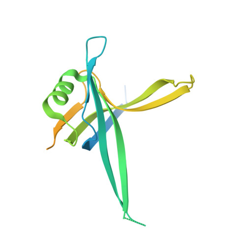 | |
UniProt | |||||
Find proteins for A0A0H3LX24 (Bartonella henselae (strain ATCC 49882 / DSM 28221 / CCUG 30454 / Houston 1)) Explore A0A0H3LX24 Go to UniProtKB: A0A0H3LX24 | |||||
Entity Groups | |||||
| Sequence Clusters | 30% Identity50% Identity70% Identity90% Identity95% Identity100% Identity | ||||
| UniProt Group | A0A0H3LX24 | ||||
Sequence AnnotationsExpand | |||||
| |||||
| Ligands 1 Unique | |||||
|---|---|---|---|---|---|
| ID | Chains | Name / Formula / InChI Key | 2D Diagram | 3D Interactions | |
| UNX Query on UNX | C [auth B] D [auth B] E [auth B] F [auth B] G [auth B] | UNKNOWN ATOM OR ION X |  | ||
| Length ( Å ) | Angle ( ˚ ) |
|---|---|
| a = 93.76 | α = 90 |
| b = 93.76 | β = 90 |
| c = 63.51 | γ = 120 |
| Software Name | Purpose |
|---|---|
| StructureStudio | data collection |
| PHASER | phasing |
| REFMAC | refinement |
| XDS | data reduction |
| XSCALE | data scaling |
RCSB PDB (citation) is hosted by
RCSB PDB is a member of the

