3M7D

Crystal structure of an N-terminal 44 kDA fragment of topoisomerase V in the presence of dioxane


Experimental Data Snapshot

  • Method: X-RAY DIFFRACTION
  • Resolution: 1.81 Å
  • R-Value Free: 0.220 
  • R-Value Work: 0.175 
  • R-Value Observed: 0.177 

wwPDB Validation   3D Report Full Report


This is version 1.2 of the entry. See complete history


Literature

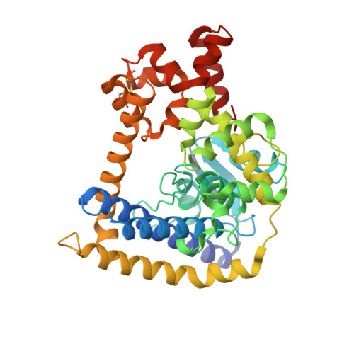
Structures of minimal catalytic fragments of topoisomerase v reveals conformational changes relevant for DNA binding.

Rajan, R.Taneja, B.Mondragon, A.

(2010) Structure 18: 829-838

  • DOI: https://doi.org/10.1016/j.str.2010.03.006
  • Primary Citation of Related Structures:  
    3M6K, 3M6Z, 3M7D, 3M7G

  • PubMed Abstract: 

    Topoisomerase V is an archaeal type I topoisomerase that is unique among topoisomerases due to presence of both topoisomerase and DNA repair activities in the same protein. It is organized as an N-terminal topoisomerase domain followed by 24 tandem helix-hairpin-helix (HhH) motifs. Structural studies have shown that the active site is buried by the (HhH) motifs. Here we show that the N-terminal domain can relax DNA in the absence of any HhH motifs and that the HhH motifs are required for stable protein-DNA complex formation. Crystal structures of various topoisomerase V fragments show changes in the relative orientation of the domains mediated by a long bent linker helix, and these movements are essential for the DNA to enter the active site. Phosphate ions bound to the protein near the active site helped model DNA in the topoisomerase domain and show how topoisomerase V may interact with DNA.


  • Organizational Affiliation

    Department of Biochemistry, Molecular Biology and Cell Biology, Northwestern University, 2205 Tech Drive, Evanston, IL 60208, USA.


Macromolecules
Find similar proteins by:  (by identity cutoff)  |  3D Structure
Entity ID: 1
MoleculeChains Sequence LengthOrganismDetailsImage
Topoisomerase V380Methanopyrus kandleri AV19Mutation(s): 0 
Gene Names: MK1436top5Topoisomerase V
UniProt
Find proteins for Q977W1 (Methanopyrus kandleri)
Explore Q977W1 
Go to UniProtKB:  Q977W1
Entity Groups  
Sequence Clusters30% Identity50% Identity70% Identity90% Identity95% Identity100% Identity
UniProt GroupQ977W1
Sequence Annotations
Expand
  • Reference Sequence
Experimental Data & Validation

Experimental Data

  • Method: X-RAY DIFFRACTION
  • Resolution: 1.81 Å
  • R-Value Free: 0.220 
  • R-Value Work: 0.175 
  • R-Value Observed: 0.177 
  • Space Group: C 1 2 1
Unit Cell:
Length ( Å )Angle ( ˚ )
a = 104.15α = 90
b = 47.74β = 112.48
c = 81.223γ = 90
Software Package:
Software NamePurpose
AMoREphasing
PHENIXrefinement
XDSdata reduction
SCALAdata scaling

Structure Validation

View Full Validation Report


Entry History 

Deposition Data

Revision History  (Full details and data files)

  • Version 1.0: 2010-08-04
    Type: Initial release
  • Version 1.1: 2011-07-13
    Changes: Version format compliance
  • Version 1.2: 2023-09-06
    Changes: Data collection, Database references, Refinement description