3LFN

Crystal structure of CDK2 with SAR57, an aminoindazole type inhibitor


Experimental Data Snapshot

  • Method: X-RAY DIFFRACTION
  • Resolution: 2.28 Å
  • R-Value Free: 0.268 
  • R-Value Work: 0.184 
  • R-Value Observed: 0.190 

wwPDB Validation   3D Report Full Report


Ligand Structure Quality Assessment 


This is version 1.2 of the entry. See complete history


Literature

Rational design of potent GSK3beta inhibitors with selectivity for Cdk1 and Cdk2.

Lesuisse, D.Dutruc-Rosset, G.Tiraboschi, G.Dreyer, M.K.Maignan, S.Chevalier, A.Halley, F.Bertrand, P.Burgevin, M.C.Quarteronet, D.Rooney, T.

(2010) Bioorg Med Chem Lett 20: 1985-1989

  • DOI: https://doi.org/10.1016/j.bmcl.2010.01.114
  • Primary Citation of Related Structures:  
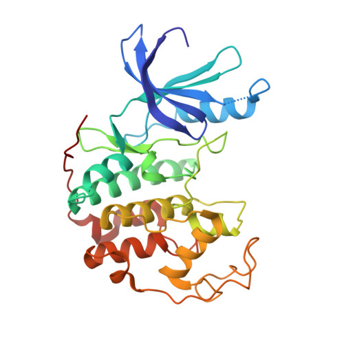
    3LAU, 3LFN, 3LFQ, 3LFS

  • PubMed Abstract: 

    From an HTS hit, a series of potent and selective inhibitors of GSK3beta have been designed based on a Cdk2-homology model and with the help of several crystal structures of the compounds within Cdk2.


  • Organizational Affiliation

    Medicinal Chemistry, 13 Quai Jules Guesde, 94300 Vitry-sur-Seine, France. dominique.lesuisse@sanofi-aventis.com


Macromolecules
Find similar proteins by:  (by identity cutoff)  |  3D Structure
Entity ID: 1
MoleculeChains Sequence LengthOrganismDetailsImage
Cell division protein kinase 2298Homo sapiensMutation(s): 0 
Gene Names: CDK2
EC: 2.7.11.22
UniProt & NIH Common Fund Data Resources
Find proteins for P24941 (Homo sapiens)
Explore P24941 
Go to UniProtKB:  P24941
PHAROS:  P24941
GTEx:  ENSG00000123374 
Entity Groups  
Sequence Clusters30% Identity50% Identity70% Identity90% Identity95% Identity100% Identity
UniProt GroupP24941
Sequence Annotations
Expand
  • Reference Sequence
Small Molecules
Ligands 1 Unique
IDChains Name / Formula / InChI Key2D Diagram3D Interactions
A27
Query on A27

Download Ideal Coordinates CCD File 
B [auth A]N-[6-(4-hydroxyphenyl)-5-phenyl-1H-indazol-3-yl]butanamide
C23 H21 N3 O2
KYYOHACAJWYNOT-UHFFFAOYSA-N
Binding Affinity Annotations 
IDSourceBinding Affinity
A27 Binding MOAD:  3LFN IC50: 3160 (nM) from 1 assay(s)
PDBBind:  3LFN IC50: 3160 (nM) from 1 assay(s)
Experimental Data & Validation

Experimental Data

  • Method: X-RAY DIFFRACTION
  • Resolution: 2.28 Å
  • R-Value Free: 0.268 
  • R-Value Work: 0.184 
  • R-Value Observed: 0.190 
  • Space Group: P 21 21 21
Unit Cell:
Length ( Å )Angle ( ˚ )
a = 53.337α = 90
b = 72.066β = 90
c = 71.439γ = 90
Software Package:
Software NamePurpose
XSCALEdata processing
TNTrefinement
PDB_EXTRACTdata extraction
XDSdata reduction
XSCALEdata scaling
BUSTERrefinement

Structure Validation

View Full Validation Report


Ligand Structure Quality Assessment 


Entry History 

Deposition Data

Revision History  (Full details and data files)

  • Version 1.0: 2010-03-02
    Type: Initial release
  • Version 1.1: 2011-07-13
    Changes: Version format compliance
  • Version 1.2: 2024-02-21
    Changes: Data collection, Database references, Derived calculations