The role of the glycan moiety on the structure-function relationships of PD-L1, type 1 ribosome-inactivating protein from P. dioica leaves
Severino, V., Chambery, A., Di Maro, A., Marasco, D., Ruggiero, A., Berisio, R., Giansanti, F., Ippoliti, R., Parente, A.(2010) Mol Biosyst 6: 570-579
- PubMed: 20174685
- DOI: https://doi.org/10.1039/b919801f
- Primary Citation of Related Structures:
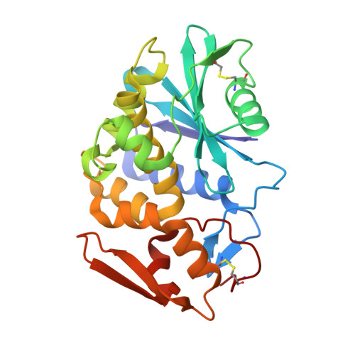
3LE7 - PubMed Abstract:
N-glycosylation is one of the major naturally occurring covalent co-translational modifications of proteins in plants, being involved in proteins structure, folding, stability and biological activity. In the present work the influence of carbohydrate moieties on the structure-function relationships of type 1 ribosome-inactivating proteins (RIPs) was investigated. To this aim, PD-Ls, RIPs isolated from Phytolacca dioica L. leaves, differing for their glycosylation degree, were used as an experimental system. In particular, comparative structural and biological analyses were performed using native and unglycosylated recombinant PD-L1, the most glycosylated P. dioica RIP isoform. The glycans influence on protein synthesis inhibition and adenine polynucleotide glycosidase activity was investigated. The interaction with adenine, the product of the de-adenylation reaction, was also investigated for native and recombinant PD-L1 by fluorescence spectroscopy. Furthermore, the crystal structure of PD-L1 in complex with adenine was determined. Our data confirm that the absence of glycan moieties did not affect the biological activity in terms of protein synthesis inhibition. However, the removal of carbohydrate chains significantly increased the deadenylation capability, likely as a consequence of the increased accessibility of substrates to the active site pocket. Furthermore, preliminary data on cellular uptake showed that all PD-L isoforms were internalized and, for the first time, that the vesicular distribution within cells could be influenced by the protein glycosylation degree.
Organizational Affiliation:
Dipartimento di Scienze della Vita, Seconda Università di Napoli, Via Vivaldi 43, I-81100 Caserta, Italy.