Crystal structure of the entire ectodomain of gp130: insights into the molecular assembly of the tall cytokine receptor complexes.
Xu, Y., Kershaw, N.J., Luo, C.S., Soo, P., Pocock, M.J., Czabotar, P.E., Hilton, D.J., Nicola, N.A., Garrett, T.P., Zhang, J.G.(2010) J Biol Chem 285: 21214-21218
- PubMed: 20489211
- DOI: https://doi.org/10.1074/jbc.C110.129502
- Primary Citation of Related Structures:
3L5H, 3L5I, 3L5J - PubMed Abstract:
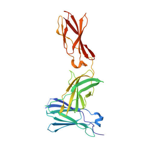
gp130 is the shared signal-transducing receptor subunit for the large and important family of interleukin 6-like cytokines. Previous x-ray structures of ligand-receptor complexes of this family lack the three membrane-proximal domains that are essential for signal transduction. Here we report the crystal structure of the entire extracellular portion of human gp130 (domains 1-6, D1-D6) at 3.6 A resolution, in an unliganded form, as well as a higher resolution structure of the membrane-proximal fibronectin type III domains (D4-D6) at 1.9 A. This represents the first atomic resolution structure of the complete ectodomain of any "tall" cytokine receptor. These structures show that other than a reorientation of the D1 domain, there is little structural change in gp130 upon ligand binding. They also reveal that the interface between the D4 and D5 domains forms an acute bend in the gp130 structure. Key residues at this interface are highly conserved across the entire tall receptor family, suggesting that this acute bend may be a common feature of these receptors. Importantly, this geometry positions the C termini of the membrane-proximal fibronectin type III domains of the tall cytokine receptors in close proximity within the transmembrane complex, favorable for receptor-associated Janus kinases to trans-phosphorylate and activate each other.
Organizational Affiliation:
Walter and Eliza Hall Institute of Medical Research, 1G Royal Parade, Parkville, Victoria 3052, Australia.