3KBC

Crystal structure of GltPh K55C-A364C mutant crosslinked with divalent mercury


Experimental Data Snapshot

  • Method: X-RAY DIFFRACTION
  • Resolution: 3.51 Å
  • R-Value Free: 0.270 
  • R-Value Work: 0.267 
  • R-Value Observed: 0.267 

wwPDB Validation   3D Report Full Report


This is version 1.3 of the entry. See complete history


Literature

Transport mechanism of a bacterial homologue of glutamate transporters.

Reyes, N.Ginter, C.Boudker, O.

(2009) Nature 462: 880-885

  • DOI: https://doi.org/10.1038/nature08616
  • Primary Citation of Related Structures:  
    3KBC

  • PubMed Abstract: 

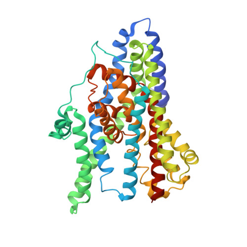
    Glutamate transporters are integral membrane proteins that catalyse a thermodynamically uphill uptake of the neurotransmitter glutamate from the synaptic cleft into the cytoplasm of glia and neuronal cells by harnessing the energy of pre-existing electrochemical gradients of ions. Crucial to the reaction is the conformational transition of the transporters between outward and inward facing states, in which the substrate binding sites are accessible from the extracellular space and the cytoplasm, respectively. Here we describe the crystal structure of a double cysteine mutant of a glutamate transporter homologue from Pyrococcus horikoshii, Glt(Ph), which is trapped in the inward facing state by cysteine crosslinking. Together with the previously determined crystal structures of Glt(Ph) in the outward facing state, the structure of the crosslinked mutant allows us to propose a molecular mechanism by which Glt(Ph) and, by analogy, mammalian glutamate transporters mediate sodium-coupled substrate uptake.


  • Organizational Affiliation

    Department of Physiology and Biophysics, Weill Cornell Medical College, 1300 York Avenue, Box 75, New York, New York 10065, USA.


Macromolecules
Find similar proteins by:  (by identity cutoff)  |  3D Structure
Entity ID: 1
MoleculeChains Sequence LengthOrganismDetailsImage
425aa long hypothetical proton glutamate symport protein
A, B, C
425Pyrococcus horikoshiiMutation(s): 10 
Gene Names: PH1295
Membrane Entity: Yes 
UniProt
Find proteins for O59010 (Pyrococcus horikoshii (strain ATCC 700860 / DSM 12428 / JCM 9974 / NBRC 100139 / OT-3))
Explore O59010 
Go to UniProtKB:  O59010
Entity Groups  
Sequence Clusters30% Identity50% Identity70% Identity90% Identity95% Identity100% Identity
UniProt GroupO59010
Sequence Annotations
Expand
  • Reference Sequence
Small Molecules
Ligands 3 Unique
IDChains Name / Formula / InChI Key2D Diagram3D Interactions
HG
Query on HG

Download Ideal Coordinates CCD File 
E [auth A],
I [auth B],
M [auth C]
MERCURY (II) ION
Hg
BQPIGGFYSBELGY-UHFFFAOYSA-N
ASP
Query on ASP

Download Ideal Coordinates CCD File 
D [auth A],
H [auth B],
L [auth C]
ASPARTIC ACID
C4 H7 N O4
CKLJMWTZIZZHCS-REOHCLBHSA-N
NA
Query on NA

Download Ideal Coordinates CCD File 
F [auth A]
G [auth A]
J [auth B]
K [auth B]
N [auth C]
F [auth A],
G [auth A],
J [auth B],
K [auth B],
N [auth C],
O [auth C]
SODIUM ION
Na
FKNQFGJONOIPTF-UHFFFAOYSA-N
Experimental Data & Validation

Experimental Data

  • Method: X-RAY DIFFRACTION
  • Resolution: 3.51 Å
  • R-Value Free: 0.270 
  • R-Value Work: 0.267 
  • R-Value Observed: 0.267 
  • Space Group: C 2 2 21
Unit Cell:
Length ( Å )Angle ( ˚ )
a = 113.142α = 90
b = 206.78β = 90
c = 205.971γ = 90
Software Package:
Software NamePurpose
REFMACrefinement
PDB_EXTRACTdata extraction
CBASSdata collection
DENZOdata reduction
SCALEPACKdata scaling
PHASERphasing

Structure Validation

View Full Validation Report


Entry History 

Deposition Data

Revision History  (Full details and data files)

  • Version 1.0: 2009-12-01
    Type: Initial release
  • Version 1.1: 2011-07-13
    Changes: Advisory, Refinement description, Version format compliance
  • Version 1.2: 2021-10-13
    Changes: Database references, Derived calculations
  • Version 1.3: 2023-09-06
    Changes: Data collection, Refinement description