Structure of a two-domain N-terminal fragment of ribosomal protein L10 from Methanococcus jannaschii reveals a specific piece of the archaeal ribosomal stalk
Kravchenko, O., Mitroshin, I., Nikonov, S., Piendl, W., Garber, M.(2010) J Mol Biol 399: 214-220
- PubMed: 20399793
- DOI: https://doi.org/10.1016/j.jmb.2010.04.017
- Primary Citation of Related Structures:
3JSY - PubMed Abstract:
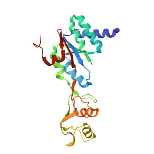
Ribosomal stalk is involved in the formation of the so-called "GTPase-associated site" and plays a key role in the interaction of ribosome with translation factors and in the control of translation accuracy. The stalk is formed by two or three copies of the L7/L12 dimer bound to the C-terminal tail of protein L10. The N-terminal domain of L10 binds to a segment of domain II of 23S rRNA near the binding site for ribosomal protein L11. The structure of bacterial L10 in complex with three L7/L12 N-terminal dimers has been determined in the isolated state, and the structure of the first third of archaeal L10 bound to domain II of 23S rRNA has been solved within the Haloarcula marismortui 50S ribosomal subunit. A close structural similarity between the RNA-binding domain of archaeal L10 and the RNA-binding domain of bacterial L10 has been demonstrated. In this work, a long RNA-binding N-terminal fragment of L10 from Methanococcus jannaschii has been isolated and crystallized. The crystal structure of this fragment (which encompasses two-thirds of the protein) has been solved at 1.6 A resolution. The model presented shows the structure of the RNA-binding domain and the structure of the adjacent domain that exist in archaeal L10 and eukaryotic P0 proteins only. Furthermore, our model incorporated into the structure of the H. marismortui 50S ribosomal subunit allows clarification of the structure of the archaeal ribosomal stalk base.
Organizational Affiliation:
Institute of Protein Research, Russian Academy of Sciences, 142290 Pushchino, Moscow Region, Russia. olesyak@rambler.ru