3IEU

Crystal Structure of ERA in Complex with GDP


Experimental Data Snapshot

  • Method: X-RAY DIFFRACTION
  • Resolution: 2.80 Å
  • R-Value Free: 0.238 
  • R-Value Work: 0.206 
  • R-Value Observed: 0.208 

wwPDB Validation   3D Report Full Report


Ligand Structure Quality Assessment 


This is version 1.4 of the entry. See complete history


Literature

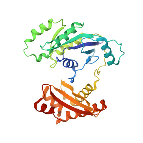
Structure of ERA in complex with the 3' end of 16S rRNA: implications for ribosome biogenesis

Tu, C.Zhou, X.Tropea, J.E.Austin, B.P.Waugh, D.S.Court, D.L.Ji, X.

(2009) Proc Natl Acad Sci U S A 106: 14843-14848

  • DOI: https://doi.org/10.1073/pnas.0904032106
  • Primary Citation of Related Structures:  
    3IEU, 3IEV

  • PubMed Abstract: 

    ERA, composed of an N-terminal GTPase domain followed by an RNA-binding KH domain, is essential for bacterial cell viability. It binds to 16S rRNA and the 30S ribosomal subunit. However, its RNA-binding site, the functional relationship between the two domains, and its role in ribosome biogenesis remain unclear. We have determined two crystal structures of ERA, a binary complex with GDP and a ternary complex with a GTP-analog and the 1531AUCACCUCCUUA1542 sequence at the 3' end of 16S rRNA. In the ternary complex, the first nine of the 12 nucleotides are recognized by the protein. We show that GTP binding is a prerequisite for RNA recognition by ERA and that RNA recognition stimulates its GTP-hydrolyzing activity. Based on these and other data, we propose a functional cycle of ERA, suggesting that the protein serves as a chaperone for processing and maturation of 16S rRNA and a checkpoint for assembly of the 30S ribosomal subunit. The AUCA sequence is highly conserved among bacteria, archaea, and eukaryotes, whereas the CCUCC, known as the anti-Shine-Dalgarno sequence, is conserved in noneukaryotes only. Therefore, these data suggest a common mechanism for a highly conserved ERA function in all three kingdoms of life by recognizing the AUCA, with a "twist" for noneukaryotic ERA proteins by also recognizing the CCUCC.


  • Organizational Affiliation

    Center for Cancer Research, National Cancer Institute, National Institutes of Health, Frederick, MD 21702, USA.


Macromolecules
Find similar proteins by:  (by identity cutoff)  |  3D Structure
Entity ID: 1
MoleculeChains Sequence LengthOrganismDetailsImage
GTP-binding protein era
A, B
301Escherichia coli K-12Mutation(s): 0 
Gene Names: b2566eraEscherichia coliJW2550rbaA
UniProt
Find proteins for P06616 (Escherichia coli (strain K12))
Explore P06616 
Go to UniProtKB:  P06616
Entity Groups  
Sequence Clusters30% Identity50% Identity70% Identity90% Identity95% Identity100% Identity
UniProt GroupP06616
Sequence Annotations
Expand
  • Reference Sequence
Small Molecules
Ligands 3 Unique
IDChains Name / Formula / InChI Key2D Diagram3D Interactions
GDP
Query on GDP

Download Ideal Coordinates CCD File 
C [auth A],
J [auth B]
GUANOSINE-5'-DIPHOSPHATE
C10 H15 N5 O11 P2
QGWNDRXFNXRZMB-UUOKFMHZSA-N
TRS
Query on TRS

Download Ideal Coordinates CCD File 
I [auth A]2-AMINO-2-HYDROXYMETHYL-PROPANE-1,3-DIOL
C4 H12 N O3
LENZDBCJOHFCAS-UHFFFAOYSA-O
SO4
Query on SO4

Download Ideal Coordinates CCD File 
D [auth A]
E [auth A]
F [auth A]
G [auth A]
H [auth A]
D [auth A],
E [auth A],
F [auth A],
G [auth A],
H [auth A],
K [auth B],
L [auth B],
M [auth B],
N [auth B],
O [auth B],
P [auth B],
Q [auth B],
R [auth B]
SULFATE ION
O4 S
QAOWNCQODCNURD-UHFFFAOYSA-L
Experimental Data & Validation

Experimental Data

  • Method: X-RAY DIFFRACTION
  • Resolution: 2.80 Å
  • R-Value Free: 0.238 
  • R-Value Work: 0.206 
  • R-Value Observed: 0.208 
  • Space Group: P 1 21 1
Unit Cell:
Length ( Å )Angle ( ˚ )
a = 86.6α = 90
b = 67.9β = 114.2
c = 87.1γ = 90
Software Package:
Software NamePurpose
PHENIXrefinement
CNSrefinement
HKL-2000data collection
HKL-2000data reduction
HKL-2000data scaling
CNSphasing

Structure Validation

View Full Validation Report


Ligand Structure Quality Assessment 


Entry History 

Deposition Data

  • Released Date: 2009-08-25 
  • Deposition Author(s): Tu, C., Ji, X.

Revision History  (Full details and data files)

  • Version 1.0: 2009-08-25
    Type: Initial release
  • Version 1.1: 2011-07-13
    Changes: Version format compliance
  • Version 1.2: 2019-07-24
    Changes: Data collection, Refinement description
  • Version 1.3: 2023-08-30
    Changes: Data collection, Database references, Derived calculations, Structure summary
  • Version 1.4: 2023-09-06
    Changes: Refinement description