3GRH

Crystal structure of escherichia coli ybhc


Experimental Data Snapshot

  • Method: X-RAY DIFFRACTION
  • Resolution: 1.70 Å
  • R-Value Free: 0.208 
  • R-Value Work: 0.193 
  • R-Value Observed: 0.193 

wwPDB Validation   3D Report Full Report


This is version 1.3 of the entry. See complete history


Literature

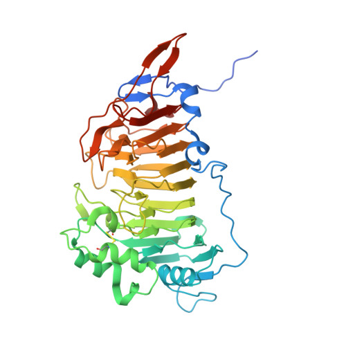
The crystal structure of the outer membrane lipoprotein YbhC from Escherichia coli sheds new light on the phylogeny of carbohydrate esterase family 8.

Eklof, J.M.Tan, T.C.Divne, C.Brumer, H.

(2009) Proteins 76: 1029-1036


Macromolecules
Find similar proteins by:  (by identity cutoff)  |  3D Structure
Entity ID: 1
MoleculeChains Sequence LengthOrganismDetailsImage
Acyl-CoA thioester hydrolase ybgC422Escherichia coli K-12Mutation(s): 0 
Gene Names: b0772JW0755ybhC
UniProt
Find proteins for P46130 (Escherichia coli (strain K12))
Explore P46130 
Go to UniProtKB:  P46130
Entity Groups  
Sequence Clusters30% Identity50% Identity70% Identity90% Identity95% Identity100% Identity
UniProt GroupP46130
Sequence Annotations
Expand
  • Reference Sequence
Experimental Data & Validation

Experimental Data

  • Method: X-RAY DIFFRACTION
  • Resolution: 1.70 Å
  • R-Value Free: 0.208 
  • R-Value Work: 0.193 
  • R-Value Observed: 0.193 
  • Space Group: P 31 2 1
Unit Cell:
Length ( Å )Angle ( ˚ )
a = 79.2α = 90
b = 79.2β = 90
c = 159.65γ = 120
Software Package:
Software NamePurpose
ProDCdata collection
SHELXSphasing
REFMACrefinement
XDSdata reduction
XSCALEdata scaling

Structure Validation

View Full Validation Report


Entry History 

Deposition Data

Revision History  (Full details and data files)

  • Version 1.0: 2009-04-07
    Type: Initial release
  • Version 1.1: 2011-07-13
    Changes: Advisory, Version format compliance
  • Version 1.2: 2018-05-23
    Changes: Data collection
  • Version 1.3: 2019-12-11
    Changes: Data collection, Database references