3GMH

Crystal Structure of the Mad2 Dimer


Experimental Data Snapshot

  • Method: X-RAY DIFFRACTION
  • Resolution: 3.95 Å
  • R-Value Free: 0.251 
  • R-Value Work: 0.218 
  • R-Value Observed: 0.220 

wwPDB Validation   3D Report Full Report


This is version 1.4 of the entry. See complete history


Literature

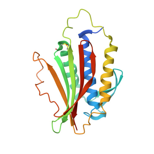
Structure of an intermediate conformer of the spindle checkpoint protein Mad2.

Hara, M.Ozkan, E.Sun, H.Yu, H.Luo, X.

(2015) Proc Natl Acad Sci U S A 112: 11252-11257

  • DOI: https://doi.org/10.1073/pnas.1512197112
  • Primary Citation of Related Structures:  
    3GMH

  • PubMed Abstract: 

    The spindle checkpoint senses unattached kinetochores during prometaphase and inhibits the anaphase-promoting complex or cyclosome (APC/C), thus ensuring accurate chromosome segregation. The checkpoint protein mitotic arrest deficient 2 (Mad2) is an unusual protein with multiple folded states. Mad2 adopts the closed conformation (C-Mad2) in a Mad1-Mad2 core complex. In mitosis, kinetochore-bound Mad1-C-Mad2 recruits latent, open Mad2 (O-Mad2) from the cytosol and converts it to an intermediate conformer (I-Mad2), which can then bind and inhibit the APC/C activator cell division cycle 20 (Cdc20) as C-Mad2. Here, we report the crystal structure and NMR analysis of I-Mad2 bound to C-Mad2. Although I-Mad2 retains the O-Mad2 fold in crystal and in solution, its core structural elements undergo discernible rigid-body movements and more closely resemble C-Mad2. Residues exhibiting methyl chemical shift changes in I-Mad2 form a contiguous, interior network that connects its C-Mad2-binding site to the conformationally malleable C-terminal region. Mutations of residues at the I-Mad2-C-Mad2 interface hinder I-Mad2 formation and impede the structural transition of Mad2. Our study provides insight into the conformational activation of Mad2 and establishes the basis of allosteric communication between two distal sites in Mad2.


  • Organizational Affiliation

    Department of Pharmacology, University of Texas Southwestern Medical Center, Dallas, TX 75390;


Macromolecules
Find similar proteins by:  (by identity cutoff)  |  3D Structure
Entity ID: 1
MoleculeChains Sequence LengthOrganismDetailsImage
Mitotic spindle assembly checkpoint protein MAD2A
A, B, C, D, E
A, B, C, D, E, F, G, H, I, J, K, L
207Homo sapiensMutation(s): 0 
Gene Names: MAD2MAD2AMAD2L1
UniProt & NIH Common Fund Data Resources
Find proteins for Q13257 (Homo sapiens)
Explore Q13257 
Go to UniProtKB:  Q13257
PHAROS:  Q13257
GTEx:  ENSG00000164109 
Entity Groups  
Sequence Clusters30% Identity50% Identity70% Identity90% Identity95% Identity100% Identity
UniProt GroupQ13257
Sequence Annotations
Expand
  • Reference Sequence
Small Molecules
Ligands 1 Unique
IDChains Name / Formula / InChI Key2D Diagram3D Interactions
SO4
Query on SO4

Download Ideal Coordinates CCD File 
M [auth A]
N [auth A]
O [auth B]
P [auth C]
Q [auth C]
M [auth A],
N [auth A],
O [auth B],
P [auth C],
Q [auth C],
R [auth D],
S [auth E],
T [auth G],
U [auth H],
V [auth I],
W [auth K],
X [auth L]
SULFATE ION
O4 S
QAOWNCQODCNURD-UHFFFAOYSA-L
Experimental Data & Validation

Experimental Data

  • Method: X-RAY DIFFRACTION
  • Resolution: 3.95 Å
  • R-Value Free: 0.251 
  • R-Value Work: 0.218 
  • R-Value Observed: 0.220 
  • Space Group: P 1 21 1
Unit Cell:
Length ( Å )Angle ( ˚ )
a = 124.829α = 90
b = 104.918β = 97.57
c = 157.746γ = 90
Software Package:
Software NamePurpose
HKL-2000data collection
PHASERphasing
PHENIXrefinement
HKL-2000data reduction
HKL-2000data scaling

Structure Validation

View Full Validation Report


Entry History 

Deposition Data

Revision History  (Full details and data files)

  • Version 1.0: 2010-11-17
    Type: Initial release
  • Version 1.1: 2011-07-13
    Changes: Version format compliance
  • Version 1.2: 2015-09-02
    Changes: Database references
  • Version 1.3: 2015-10-07
    Changes: Database references
  • Version 1.4: 2023-09-06
    Changes: Data collection, Database references, Derived calculations, Refinement description