3ES6

Crystal structure of the novel complex formed between Zinc 2-glycoprotein (ZAG) and Prolactin inducible protein (PIP) from human seminal plasma


Experimental Data Snapshot

  • Method: X-RAY DIFFRACTION
  • Resolution: 3.23 Å
  • R-Value Free: 0.240 
  • R-Value Work: 0.196 
  • R-Value Observed: 0.200 

wwPDB Validation   3D Report Full Report


This is version 3.1 of the entry. See complete history


Literature

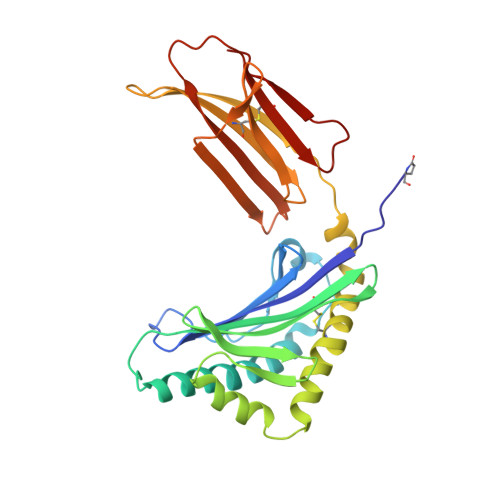
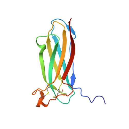
Crystal structure of the novel complex formed between zinc alpha2-glycoprotein (ZAG) and prolactin-inducible protein (PIP) from human seminal plasma

Hassan, M.I.Bilgrami, S.Kumar, V.Singh, N.Yadav, S.Kaur, P.Singh, T.P.

(2008) J Mol Biol 384: 663-672

  • DOI: https://doi.org/10.1016/j.jmb.2008.09.072
  • Primary Citation of Related Structures:  
    3ES6

  • PubMed Abstract: 

    This is the first report on the formation of a complex between zinc alpha2-glycoprotein (ZAG) and prolactin-inducible protein (PIP). The complex was purified from human seminal plasma and crystallized using 20% polyethylene glycol 9000 and 5% hexaethylene glycol. The structure of the complex has been determined using X-ray crystallographic method and refined to an R(cryst) of 0.199 (R(free)=0.239). The structure of ZAG is broadly similar to the structure of serum ZAG. The scaffolding of PIP consists of seven beta-strands that are organized in the form of two antiparallel beta-pleated sheets, resulting in the formation of a sandwiched beta-sheet. The amino acid sequence of PIP contains one potential N-glycosylation site at Asn77, and the same is found glycosylated with four sugar residues. The structure of the complex shows that the beta-structure of PIP is ideally aligned with the beta-structure of domain alpha3 of ZAG to form a long interface between two proteins. The proximal beta-strands at the long interface are arranged in an antiparallel manner. There are 12 hydrogen bonds and three salt bridges between ZAG and PIP. At the two ends of vertical interface, two salt bridges are formed between pairs of Lys41-Asp233 and Lys68-Glu229. On the perpendicular interface involving alpha1-alpha2 domains of ZAG and a loop of PIP, another salt bridge is formed. The internal space at the corner of the L-shaped structure is filled with solvent molecules including a carbonate ion. The overall buried area in the complex is approximately 914 A(2), which is considerably higher than the 660 A(2) reported for the class I major histocompatibility complex structures.


  • Organizational Affiliation

    Department of Biophysics, All India Institute of Medical Sciences, New Delhi 110029, India.


Macromolecules
Find similar proteins by:  (by identity cutoff)  |  3D Structure
Entity ID: 1
MoleculeChains Sequence LengthOrganismDetailsImage
Zinc-alpha-2-glycoprotein278Homo sapiensMutation(s): 0 
UniProt & NIH Common Fund Data Resources
Find proteins for P25311 (Homo sapiens)
Explore P25311 
Go to UniProtKB:  P25311
PHAROS:  P25311
GTEx:  ENSG00000160862 
Entity Groups  
Sequence Clusters30% Identity50% Identity70% Identity90% Identity95% Identity100% Identity
UniProt GroupP25311
Sequence Annotations
Expand
  • Reference Sequence
Find similar proteins by:  (by identity cutoff)  |  3D Structure
Entity ID: 2
MoleculeChains Sequence LengthOrganismDetailsImage
Prolactin-inducible protein118Homo sapiensMutation(s): 0 
UniProt & NIH Common Fund Data Resources
Find proteins for P12273 (Homo sapiens)
Explore P12273 
Go to UniProtKB:  P12273
PHAROS:  P12273
GTEx:  ENSG00000159763 
Entity Groups  
Sequence Clusters30% Identity50% Identity70% Identity90% Identity95% Identity100% Identity
UniProt GroupP12273
Sequence Annotations
Expand
  • Reference Sequence
Oligosaccharides

Help

Entity ID: 3
MoleculeChains Length2D Diagram Glycosylation3D Interactions
2-acetamido-2-deoxy-alpha-D-glucopyranose-(1-2)-alpha-D-mannopyranose-(1-3)-beta-D-mannopyranose-(1-4)-2-acetamido-2-deoxy-beta-D-glucopyranose-(1-4)-2-acetamido-2-deoxy-beta-D-glucopyranose
C
5N-Glycosylation
Glycosylation Resources
GlyTouCan:  G96957PS
GlyCosmos:  G96957PS
Entity ID: 4
MoleculeChains Length2D Diagram Glycosylation3D Interactions
alpha-D-mannopyranose-(1-3)-beta-D-mannopyranose-(1-4)-2-acetamido-2-deoxy-beta-D-glucopyranose-(1-4)-2-acetamido-2-deoxy-beta-D-glucopyranose
D
4N-Glycosylation
Glycosylation Resources
GlyTouCan:  G81315DD
GlyCosmos:  G81315DD
GlyGen:  G81315DD
Small Molecules
Modified Residues  1 Unique
IDChains TypeFormula2D DiagramParent
PCA
Query on PCA
A
L-PEPTIDE LINKINGC5 H7 N O3GLN
Experimental Data & Validation

Experimental Data

  • Method: X-RAY DIFFRACTION
  • Resolution: 3.23 Å
  • R-Value Free: 0.240 
  • R-Value Work: 0.196 
  • R-Value Observed: 0.200 
  • Space Group: P 4 21 2
Unit Cell:
Length ( Å )Angle ( ˚ )
a = 132.298α = 90
b = 132.298β = 90
c = 73.752γ = 90
Software Package:
Software NamePurpose
MAR345dtbdata collection
AMoREphasing
REFMACrefinement
DENZOdata reduction
SCALEPACKdata scaling

Structure Validation

View Full Validation Report


Entry History 

Deposition Data

Revision History  (Full details and data files)

  • Version 1.0: 2008-10-28
    Type: Initial release
  • Version 1.1: 2011-07-13
    Changes: Non-polymer description, Version format compliance
  • Version 2.0: 2019-12-25
    Changes: Database references, Derived calculations, Polymer sequence
  • Version 3.0: 2020-07-29
    Type: Remediation
    Reason: Carbohydrate remediation
    Changes: Advisory, Atomic model, Data collection, Derived calculations, Structure summary
  • Version 3.1: 2023-11-01
    Changes: Data collection, Database references, Derived calculations, Refinement description, Structure summary