Structural basis for induced formation of the inflammatory mediator prostaglandin E2
Jegerschold, C., Pawelzik, S.C., Purhonen, P., Bhakat, P., Gheorghe, K.R., Gyobu, N., Mitsuoka, K., Morgenstern, R., Jakobsson, P.J., Hebert, H.(2008) Proc Natl Acad Sci U S A 105: 11110-11115
- PubMed: 18682561
- DOI: https://doi.org/10.1073/pnas.0802894105
- Primary Citation of Related Structures:
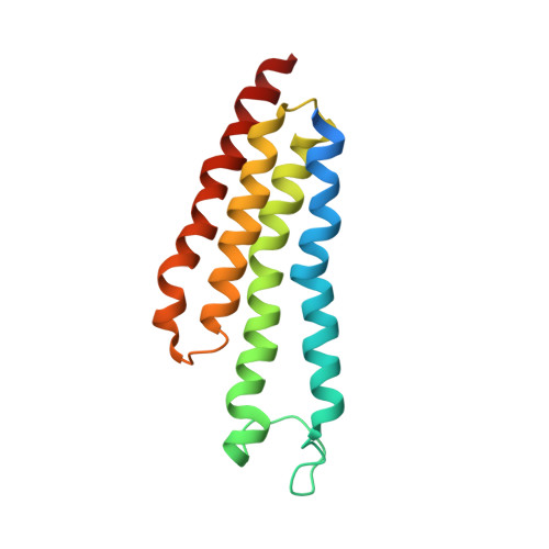
3DWW - PubMed Abstract:
Prostaglandins (PG) are bioactive lipids produced from arachidonic acid via the action of cyclooxygenases and terminal PG synthases. Microsomal prostaglandin E synthase 1 (MPGES1) constitutes an inducible glutathione-dependent integral membrane protein that catalyzes the oxidoreduction of cyclooxygenase derived PGH(2) into PGE(2). MPGES1 has been implicated in a number of human diseases or pathological conditions, such as rheumatoid arthritis, fever, and pain, and is therefore regarded as a primary target for development of novel antiinflammatory drugs. To provide a structural basis for insight in the catalytic mechanism, we determined the structure of MPGES1 in complex with glutathione by electron crystallography from 2D crystals induced in the presence of phospholipids. Together with results from site-directed mutagenesis and activity measurements, we can thereby demonstrate the role of specific amino acid residues. Glutathione is found to bind in a U-shaped conformation at the interface between subunits in the protein trimer. It is exposed to a site facing the lipid bilayer, which forms the specific environment for the oxidoreduction of PGH(2) to PGE(2) after displacement of the cytoplasmic half of the N-terminal transmembrane helix. Hence, insight into the dynamic behavior of MPGES1 and homologous membrane proteins in inflammation and detoxification is provided.
Organizational Affiliation:
Department of Biosciences and Nutrition, Karolinska Institutet and School of Technology and Health, Royal Institute of Technology, Novum, S-141 57 Huddinge, Sweden. caroline.jegerschold@ki.se