3D93

Apo Human carbonic anhydrase II bound with substrate carbon dioxide


Experimental Data Snapshot

  • Method: X-RAY DIFFRACTION
  • Resolution: 1.10 Å
  • R-Value Free: 0.139 
  • R-Value Work: 0.104 
  • R-Value Observed: 0.104 

wwPDB Validation   3D Report Full Report


This is version 1.3 of the entry. See complete history


Literature

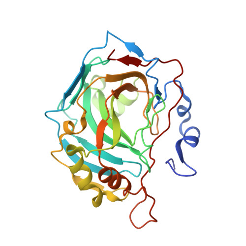
Entrapment of carbon dioxide in the active site of carbonic anhydrase II

Domsic, J.F.Avvaru, B.S.Kim, C.U.Gruner, S.M.Agbandje-McKenna, M.Silverman, D.N.McKenna, R.

(2008) J Biol Chem 283: 30766-30771

  • DOI: https://doi.org/10.1074/jbc.M805353200
  • Primary Citation of Related Structures:  
    3D92, 3D93

  • PubMed Abstract: 

    The visualization at near atomic resolution of transient substrates in the active site of enzymes is fundamental to fully understanding their mechanism of action. Here we show the application of using CO(2)-pressurized, cryo-cooled crystals to capture the first step of CO(2) hydration catalyzed by the zinc-metalloenzyme human carbonic anhydrase II, the binding of substrate CO(2), for both the holo and the apo (without zinc) enzyme to 1.1A resolution. Until now, the feasibility of such a study was thought to be technically too challenging because of the low solubility of CO(2) and the fast turnover to bicarbonate by the enzyme (Liang, J. Y., and Lipscomb, W. N. (1990) Proc. Natl. Acad. Sci. U. S. A. 87, 3675-3679). These structures provide insight into the long hypothesized binding of CO(2) in a hydrophobic pocket at the active site and demonstrate that the zinc does not play a critical role in the binding or orientation of CO(2). This method may also have a much broader implication for the study of other enzymes for which CO(2) is a substrate or product and for the capturing of transient substrates and revealing hydrophobic pockets in proteins.


  • Organizational Affiliation

    Department of Biochemistry and Molecular Biology, University of Florida, Gainesville, Florida 32610, USA.


Macromolecules
Find similar proteins by:  (by identity cutoff)  |  3D Structure
Entity ID: 1
MoleculeChains Sequence LengthOrganismDetailsImage
carbonic anhydrase II260Homo sapiensMutation(s): 0 
Gene Names: HCA2
EC: 4.2.1.1
UniProt & NIH Common Fund Data Resources
Find proteins for P00918 (Homo sapiens)
Explore P00918 
Go to UniProtKB:  P00918
PHAROS:  P00918
GTEx:  ENSG00000104267 
Entity Groups  
Sequence Clusters30% Identity50% Identity70% Identity90% Identity95% Identity100% Identity
UniProt GroupP00918
Sequence Annotations
Expand
  • Reference Sequence
Experimental Data & Validation

Experimental Data

  • Method: X-RAY DIFFRACTION
  • Resolution: 1.10 Å
  • R-Value Free: 0.139 
  • R-Value Work: 0.104 
  • R-Value Observed: 0.104 
  • Space Group: P 1 21 1
Unit Cell:
Length ( Å )Angle ( ˚ )
a = 42.215α = 90
b = 41.514β = 104.15
c = 72.346γ = 90
Software Package:
Software NamePurpose
DENZOdata reduction
SCALEPACKdata scaling
SHELXrefinement
PDB_EXTRACTdata extraction
ADSCdata collection
SHELXL-97refinement

Structure Validation

View Full Validation Report


Entry History 

Deposition Data

Revision History  (Full details and data files)

  • Version 1.0: 2008-09-02
    Type: Initial release
  • Version 1.1: 2011-07-13
    Changes: Non-polymer description, Version format compliance
  • Version 1.2: 2017-10-25
    Changes: Refinement description
  • Version 1.3: 2023-08-30
    Changes: Data collection, Database references, Derived calculations, Refinement description