3D5Z

Crystal Structure Analysis of 1,5-alpha-arabinanase catalytic mutant (AbnBE201A) complexed to arabinotriose


Experimental Data Snapshot

  • Method: X-RAY DIFFRACTION
  • Resolution: 1.90 Å
  • R-Value Free: 0.236 
  • R-Value Work: 0.192 
  • R-Value Observed: 0.192 

wwPDB Validation   3D Report Full Report


This is version 2.2 of the entry. See complete history


Literature

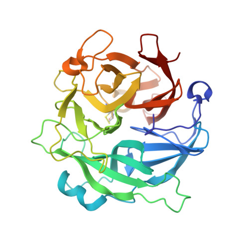
Crystal structure of an inverting GH 43 1,5-alpha-L-arabinanase from Geobacillus stearothermophilus complexed with its substrate

Alhassid, A.Ben-David, A.Tabachnikov, O.Libster, D.Naveh, E.Zolotnitsky, G.Shoham, Y.Shoham, G.

(2009) Biochem J 422: 73-82

  • DOI: https://doi.org/10.1042/BJ20090180
  • Primary Citation of Related Structures:  
    3CU9, 3D5Y, 3D5Z, 3D60, 3D61

  • PubMed Abstract: 

    Arabinanases are glycosidases that hydrolyse alpha-(1-->5)- arabinofuranosidic linkages found in the backbone of the pectic polysaccharide arabinan. Here we describe the biochemical characterization and the enzyme-substrate crystal structure of an inverting family 43 arabinanase from Geobacillus stearothermophilus T-6 (AbnB). Based on viscosity and reducing power measurements, and based on product analysis for the hydrolysis of linear arabinan by AbnB, the enzyme works in an endo mode of action. Isothermal titration calorimetry studies of a catalytic mutant with various arabino-oligosaccharides suggested that the enzyme active site can accommodate at least five arabinose units. The crystal structure of AbnB was determined at 1.06 A (1 A=0.1 nm) resolution, revealing a single five-bladed-beta-propeller fold domain. Co-crystallization of catalytic mutants of the enzyme with different substrates allowed us to obtain complex structures of AbnBE201A with arabinotriose and AbnBD147A with arabinobiose. Based on the crystal structures of AbnB together with its substrates, the position of the three catalytic carboxylates: Asp27, the general base; Glu201, the general acid; and Asp147, the pKa modulator, is in agreement with their putative catalytic roles. In the complex structure of AbnBE201A with arabinotriose, a single water molecule is located 2.8 A from Asp27 and 3.7 A from the anomeric carbon. The position of this water molecule is kept via hydrogen bonding with a conserved tyrosine (Tyr229) that is 2.6 A distant from it. The location of this molecule suggests that it can function as the catalytic water molecule in the hydrolysis reaction, resulting in the inversion of the anomeric configuration of the product.


  • Organizational Affiliation

    Institute of Chemistry, The Hebrew University of Jerusalem, Jerusalem, Israel.


Macromolecules
Find similar proteins by:  (by identity cutoff)  |  3D Structure
Entity ID: 1
MoleculeChains Sequence LengthOrganismDetailsImage
Intracellular arabinanase314Geobacillus stearothermophilusMutation(s): 1 
Gene Names: abn
EC: 3.2.1.99
UniProt
Find proteins for B3EYM8 (Geobacillus stearothermophilus)
Explore B3EYM8 
Go to UniProtKB:  B3EYM8
Entity Groups  
Sequence Clusters30% Identity50% Identity70% Identity90% Identity95% Identity100% Identity
UniProt GroupB3EYM8
Sequence Annotations
Expand
  • Reference Sequence
Oligosaccharides

Help

Entity ID: 2
MoleculeChains Length2D Diagram Glycosylation3D Interactions
alpha-L-arabinofuranose-(1-5)-alpha-L-arabinofuranose-(1-5)-beta-L-arabinofuranose
B
3N/A
Glycosylation Resources
GlyTouCan:  G42352CC
GlyCosmos:  G42352CC
Small Molecules
Ligands 1 Unique
IDChains Name / Formula / InChI Key2D Diagram3D Interactions
CA
Query on CA

Download Ideal Coordinates CCD File 
C [auth A]CALCIUM ION
Ca
BHPQYMZQTOCNFJ-UHFFFAOYSA-N
Experimental Data & Validation

Experimental Data

  • Method: X-RAY DIFFRACTION
  • Resolution: 1.90 Å
  • R-Value Free: 0.236 
  • R-Value Work: 0.192 
  • R-Value Observed: 0.192 
  • Space Group: C 2 2 21
Unit Cell:
Length ( Å )Angle ( ˚ )
a = 84.682α = 90
b = 88.831β = 90
c = 75.265γ = 90
Software Package:
Software NamePurpose
CNSrefinement
DENZOdata reduction
SCALEPACKdata scaling
CNSphasing

Structure Validation

View Full Validation Report


Entry History 

Deposition Data

Revision History  (Full details and data files)

  • Version 1.0: 2009-04-21
    Type: Initial release
  • Version 1.1: 2011-07-13
    Changes: Version format compliance
  • Version 1.2: 2017-10-25
    Changes: Refinement description
  • Version 2.0: 2020-07-29
    Type: Remediation
    Reason: Carbohydrate remediation
    Changes: Atomic model, Data collection, Derived calculations, Structure summary
  • Version 2.1: 2021-11-10
    Changes: Database references, Derived calculations, Structure summary
  • Version 2.2: 2023-11-01
    Changes: Data collection, Refinement description