3D3J

Crystal structure of human Edc3p


Experimental Data Snapshot

  • Method: X-RAY DIFFRACTION
  • Resolution: 2.80 Å
  • R-Value Free: 0.291 
  • R-Value Work: 0.229 
  • R-Value Observed: 0.232 

wwPDB Validation   3D Report Full Report


This is version 1.2 of the entry. See complete history


Literature

Crystal structure of human Edc3 and its functional implications

Ling, S.H.M.Decker, C.J.Walsh, M.A.She, M.Parker, R.Song, H.

(2008) Mol Cell Biol 28: 5965-5976

  • DOI: https://doi.org/10.1128/MCB.00761-08
  • Primary Citation of Related Structures:  
    3D3J, 3D3K

  • PubMed Abstract: 

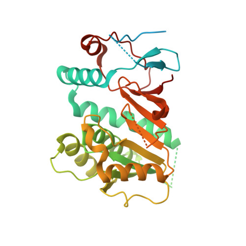
    Edc3 is an enhancer of decapping and serves as a scaffold that aggregates mRNA ribonucleoproteins together for P-body formation. Edc3 forms a network of interactions with the components of the mRNA decapping machinery and has a modular domain architecture consisting of an N-terminal Lsm domain, a central FDF domain, and a C-terminal YjeF-N domain. We have determined the crystal structure of the N-terminally truncated human Edc3 at a resolution of 2.2 A. The structure reveals that the YjeF-N domain of Edc3 possesses a divergent Rossmann fold topology that forms a dimer, which is supported by sedimentation velocity and sedimentation equilibrium analysis in solution. The dimerization interface of Edc3 is highly conserved in eukaryotes despite the overall low sequence homology across species. Structure-based site-directed mutagenesis revealed dimerization is required for efficient RNA binding, P-body formation, and likely for regulating the yeast Rps28B mRNA as well, suggesting that the dimeric form of Edc3 is a structural and functional unit in mRNA degradation.


  • Organizational Affiliation

    Laboratory of Macromolecular Structure, Institute of Molecular and Cell Biology, 61 Biopolis Drive, Singapore 138673, Singapore.


Macromolecules
Find similar proteins by:  (by identity cutoff)  |  3D Structure
Entity ID: 1
MoleculeChains Sequence LengthOrganismDetailsImage
Enhancer of mRNA-decapping protein 3306Homo sapiensMutation(s): 0 
Gene Names: EDC3
UniProt & NIH Common Fund Data Resources
Find proteins for Q96F86 (Homo sapiens)
Explore Q96F86 
Go to UniProtKB:  Q96F86
PHAROS:  Q96F86
GTEx:  ENSG00000179151 
Entity Groups  
Sequence Clusters30% Identity50% Identity70% Identity90% Identity95% Identity100% Identity
UniProt GroupQ96F86
Sequence Annotations
Expand
  • Reference Sequence
Experimental Data & Validation

Experimental Data

  • Method: X-RAY DIFFRACTION
  • Resolution: 2.80 Å
  • R-Value Free: 0.291 
  • R-Value Work: 0.229 
  • R-Value Observed: 0.232 
  • Space Group: I 4 2 2
Unit Cell:
Length ( Å )Angle ( ˚ )
a = 119.741α = 90
b = 119.741β = 90
c = 93.923γ = 90
Software Package:
Software NamePurpose
REFMACrefinement
MOSFLMdata reduction
SCALAdata scaling
PHASERphasing

Structure Validation

View Full Validation Report


Entry History 

Deposition Data

  • Released Date: 2008-08-26 
  • Deposition Author(s): Ling, S.H.M.

Revision History  (Full details and data files)

  • Version 1.0: 2008-08-26
    Type: Initial release
  • Version 1.1: 2011-07-13
    Changes: Advisory, Version format compliance
  • Version 1.2: 2023-11-01
    Changes: Data collection, Database references, Refinement description