3BZM

Crystal Structure of Open form of Menaquinone-Specific Isochorismate Synthase, MenF


Experimental Data Snapshot

  • Method: X-RAY DIFFRACTION
  • Resolution: 1.95 Å
  • R-Value Free: 0.308 
  • R-Value Work: 0.245 
  • R-Value Observed: 0.248 

wwPDB Validation   3D Report Full Report


This is version 1.2 of the entry. See complete history


Literature

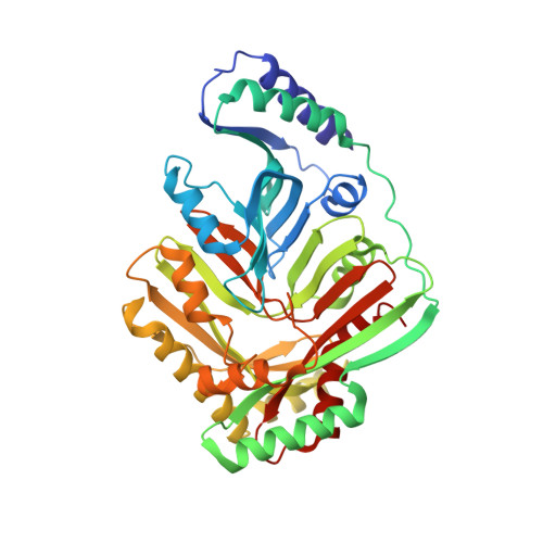
Structure of isochorismate synthase in complex with magnesium.

Parsons, J.F.Shi, K.M.Ladner, J.E.

(2008) Acta Crystallogr D Biol Crystallogr 64: 607-610

  • DOI: https://doi.org/10.1107/S0907444908005477
  • Primary Citation of Related Structures:  
    3BZM, 3BZN

  • PubMed Abstract: 

    The electron carrier menaquinone is one of many important bacterial metabolites that are derived from the key intermediate chorismic acid. MenF, the first enzyme in the menaquinone pathway, catalyzes the isomerization of chorismate to isochorismate. Here, an improved structure of MenF in a new crystal form is presented. The structure, solved at 2.0 angstroms resolution in complex with magnesium, reveals a well defined closed active site. Existing evidence suggests that the mechanism of the reaction catalyzed by MenF involves nucleophilic attack of a water molecule on the chorismate ring. The structure reveals a well defined water molecule located in an appropriate position for activation by Lys190 and attack on the substrate.


  • Organizational Affiliation

    Center for Advanced Research in Biotechnology, University of Maryland Biotechnology Institute, USA. parsonsj@umbi.umd.edu


Macromolecules
Find similar proteins by:  (by identity cutoff)  |  3D Structure
Entity ID: 1
MoleculeChains Sequence LengthOrganismDetailsImage
Menaquinone-specific isochorismate synthase431Escherichia coliMutation(s): 0 
Gene Names: menFyfbA
EC: 5.4.4.2
UniProt
Find proteins for P38051 (Escherichia coli (strain K12))
Explore P38051 
Go to UniProtKB:  P38051
Entity Groups  
Sequence Clusters30% Identity50% Identity70% Identity90% Identity95% Identity100% Identity
UniProt GroupP38051
Sequence Annotations
Expand
  • Reference Sequence
Small Molecules
Ligands 1 Unique
IDChains Name / Formula / InChI Key2D Diagram3D Interactions
CIT
Query on CIT

Download Ideal Coordinates CCD File 
B [auth A]CITRIC ACID
C6 H8 O7
KRKNYBCHXYNGOX-UHFFFAOYSA-N
Experimental Data & Validation

Experimental Data

  • Method: X-RAY DIFFRACTION
  • Resolution: 1.95 Å
  • R-Value Free: 0.308 
  • R-Value Work: 0.245 
  • R-Value Observed: 0.248 
  • Space Group: P 32 2 1
Unit Cell:
Length ( Å )Angle ( ˚ )
a = 72.37α = 90
b = 72.37β = 90
c = 164.01γ = 120
Software Package:
Software NamePurpose
d*TREKdata scaling
PHASERphasing
REFMACrefinement
PDB_EXTRACTdata extraction
CrystalCleardata collection
d*TREKdata reduction

Structure Validation

View Full Validation Report


Entry History 

Deposition Data

Revision History  (Full details and data files)

  • Version 1.0: 2008-05-20
    Type: Initial release
  • Version 1.1: 2011-07-13
    Changes: Version format compliance
  • Version 1.2: 2024-02-21
    Changes: Data collection, Database references, Derived calculations