3BUL

E. coli I690C/G743C MetH C-terminal fragment (649-1227)


Experimental Data Snapshot

  • Method: X-RAY DIFFRACTION
  • Resolution: 2.30 Å
  • R-Value Free: 0.247 
  • R-Value Work: 0.198 
  • R-Value Observed: 0.200 

wwPDB Validation   3D Report Full Report


Ligand Structure Quality Assessment 


This is version 1.4 of the entry. See complete history


Literature

A disulfide-stabilized conformer of methionine synthase reveals an unexpected role for the histidine ligand of the cobalamin cofactor.

Datta, S.Koutmos, M.Pattridge, K.A.Ludwig, M.L.Matthews, R.G.

(2008) Proc Natl Acad Sci U S A 105: 4115-4120

  • DOI: https://doi.org/10.1073/pnas.0800329105
  • Primary Citation of Related Structures:  
    3BUL

  • PubMed Abstract: 

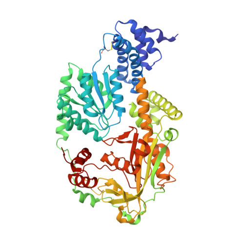
    B(12)-dependent methionine synthase (MetH) from Escherichia coli is a large modular protein that is alternately methylated by methyltetrahydrofolate to form methylcobalamin and demethylated by homocysteine to form cob(I)alamin. Major domain rearrangements are required to allow cobalamin to react with three different substrates: homocysteine, methyltetrahydrofolate, and S-adenosyl-l-methionine (AdoMet). These same rearrangements appear to preclude crystallization of the wild-type enzyme. Disulfide cross-linking was used to lock a C-terminal fragment of the enzyme into a unique conformation. Cysteine point mutations were introduced at Ile-690 and Gly-743. These cysteine residues span the cap and the cobalamin-binding module and form a cross-link that reduces the conformational space accessed by the enzyme, facilitating protein crystallization. Here, we describe an x-ray structure of the mutant fragment in the reactivation conformation; this conformation enables the transfer of a methyl group from AdoMet to the cobalamin cofactor. In the structure, the axial ligand to the cobalamin, His-759, dissociates from the cobalamin and forms intermodular contacts with residues in the AdoMet-binding module. This unanticipated intermodular interaction is expected to play a major role in controlling the distribution of conformers required for the catalytic and the reactivation cycles of the enzyme.


  • Organizational Affiliation

    Life Sciences Institute, Biophysics Research Division, and Department of Biological Chemistry, University of Michigan, Ann Arbor, MI 48109-2216, USA.


Macromolecules
Find similar proteins by:  (by identity cutoff)  |  3D Structure
Entity ID: 1
MoleculeChains Sequence LengthOrganismDetailsImage
Methionine synthase579Escherichia coliMutation(s): 2 
Gene Names: metH
EC: 2.1.1.13
UniProt
Find proteins for P13009 (Escherichia coli (strain K12))
Explore P13009 
Go to UniProtKB:  P13009
Entity Groups  
Sequence Clusters30% Identity50% Identity70% Identity90% Identity95% Identity100% Identity
UniProt GroupP13009
Sequence Annotations
Expand
  • Reference Sequence
Small Molecules
Ligands 1 Unique
IDChains Name / Formula / InChI Key2D Diagram3D Interactions
B12
Query on B12

Download Ideal Coordinates CCD File 
B [auth A]COBALAMIN
C62 H89 Co N13 O14 P
LKVIQTCSMMVGFU-DWSMJLPVSA-N
Experimental Data & Validation

Experimental Data

  • Method: X-RAY DIFFRACTION
  • Resolution: 2.30 Å
  • R-Value Free: 0.247 
  • R-Value Work: 0.198 
  • R-Value Observed: 0.200 
  • Space Group: P 43 21 2
Unit Cell:
Length ( Å )Angle ( ˚ )
a = 106.529α = 90
b = 106.529β = 90
c = 137.15γ = 90
Software Package:
Software NamePurpose
XSCALEdata scaling
EPMRphasing
REFMACrefinement
PDB_EXTRACTdata extraction
Blu-Icedata collection
XDSdata reduction

Structure Validation

View Full Validation Report


Ligand Structure Quality Assessment 


Entry History 

Deposition Data

Revision History  (Full details and data files)

  • Version 1.0: 2008-04-01
    Type: Initial release
  • Version 1.1: 2011-07-13
    Changes: Version format compliance
  • Version 1.2: 2012-10-24
    Changes: Non-polymer description
  • Version 1.3: 2021-10-20
    Changes: Database references, Derived calculations
  • Version 1.4: 2023-08-30
    Changes: Data collection, Refinement description