3BFQ

Crystal structure of truncated FimG (FimGt) in complex with the donor strand peptide of FimF (DSF)


Experimental Data Snapshot

  • Method: X-RAY DIFFRACTION
  • Resolution: 1.34 Å
  • R-Value Free: 0.179 
  • R-Value Observed: 0.132 

wwPDB Validation   3D Report Full Report


This is version 1.2 of the entry. See complete history


Literature

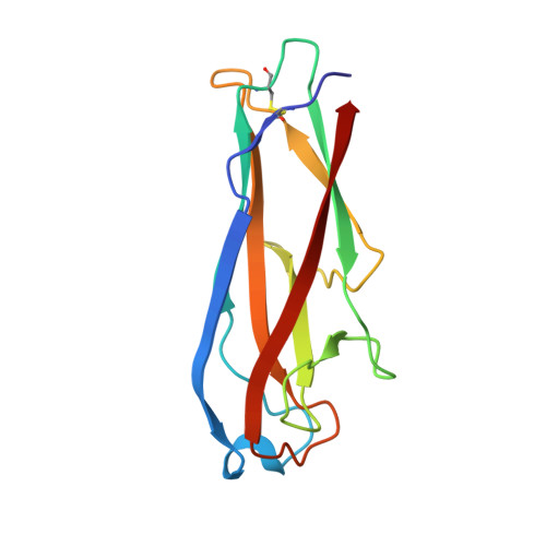
Infinite Kinetic Stability against Dissociation of Supramolecular Protein Complexes through Donor Strand Complementation

Puorger, C.Eidam, O.Capitani, G.Erilov, D.Grutter, M.G.Glockshuber, R.

(2008) Structure 16: 631-642

  • DOI: https://doi.org/10.1016/j.str.2008.01.013
  • Primary Citation of Related Structures:  
    3BFQ, 3BFW

  • PubMed Abstract: 

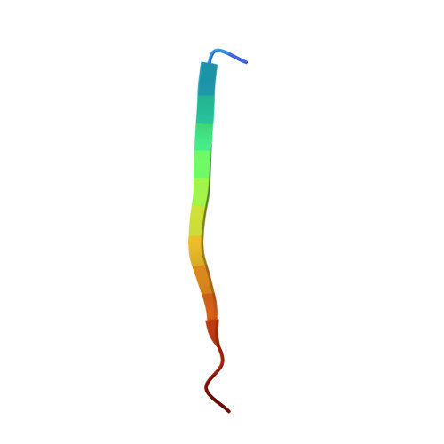
    Adhesive type 1 pili from uropathogenic Escherichia coli strains are heat and denaturant resistant, filamentous protein complexes. Individual pilus subunits associate through "donor strand complementation," whereby the incomplete immunoglobulin-like fold of each subunit is completed by the N-terminal extension of a neighboring subunit. We show that antiparallel donor strand insertion generally causes nonequilibrium behavior in protein folding and extreme activation energy barriers for dissociation of subunit-subunit complexes. We identify the most kinetically stable, noncovalent protein complex known to date. The complex between the pilus subunit FimG and the donor strand peptide of the subunit FimF shows an extrapolated dissociation half-life of 3 x 10(9) years. The 15 residue peptide forms ideal intermolecular beta sheet H-bonds with FimG over 10 residues, and its hydrophobic side chains strongly interact with the hydrophobic core of FimG. The results show that kinetic stability and nonequilibrium behavior in protein folding confers infinite stability against dissociation in extracellular protein complexes.


  • Organizational Affiliation

    ETH Zürich, Institute of Molecular Biology and Biophysics, CH-8093 Zurich, Switzerland.


Macromolecules
Find similar proteins by:  (by identity cutoff)  |  3D Structure
Entity ID: 1
MoleculeChains Sequence LengthOrganismDetailsImage
Protein fimGA [auth G]132Escherichia coli str. K-12 substr. W3110Mutation(s): 0 
Gene Names: fimG
UniProt
Find proteins for P08190 (Escherichia coli (strain K12))
Explore P08190 
Go to UniProtKB:  P08190
Entity Groups  
Sequence Clusters30% Identity50% Identity70% Identity90% Identity95% Identity100% Identity
UniProt GroupP08190
Sequence Annotations
Expand
  • Reference Sequence

Find similar proteins by:  Sequence   |   3D Structure  

Entity ID: 2
MoleculeChains Sequence LengthOrganismDetailsImage
Protein fimFB [auth F]15Escherichia coliMutation(s): 1 
UniProt
Find proteins for P08189 (Escherichia coli (strain K12))
Explore P08189 
Go to UniProtKB:  P08189
Entity Groups  
Sequence Clusters30% Identity50% Identity70% Identity90% Identity95% Identity100% Identity
UniProt GroupP08189
Sequence Annotations
Expand
  • Reference Sequence
Small Molecules
Ligands 1 Unique
IDChains Name / Formula / InChI Key2D Diagram3D Interactions
CO
Query on CO

Download Ideal Coordinates CCD File 
C [auth G]COBALT (II) ION
Co
XLJKHNWPARRRJB-UHFFFAOYSA-N
Experimental Data & Validation

Experimental Data

  • Method: X-RAY DIFFRACTION
  • Resolution: 1.34 Å
  • R-Value Free: 0.179 
  • R-Value Observed: 0.132 
  • Space Group: P 1 21 1
Unit Cell:
Length ( Å )Angle ( ˚ )
a = 48.05α = 90
b = 24.5β = 113.77
c = 54.15γ = 90
Software Package:
Software NamePurpose
SHELXrefinement
PDB_EXTRACTdata extraction
DENZOdata reduction
SCALEPACKdata scaling
SHARPphasing
SHELXL-97refinement

Structure Validation

View Full Validation Report


Entry History 

Deposition Data

Revision History  (Full details and data files)

  • Version 1.0: 2008-03-04
    Type: Initial release
  • Version 1.1: 2011-07-13
    Changes: Source and taxonomy, Version format compliance
  • Version 1.2: 2021-10-20
    Changes: Database references, Derived calculations, Source and taxonomy