3AJM

Crystal structure of programmed cell death 10 in complex with inositol 1,3,4,5-tetrakisphosphate


Experimental Data Snapshot

  • Method: X-RAY DIFFRACTION
  • Resolution: 2.30 Å
  • R-Value Free: 0.264 
  • R-Value Work: 0.213 
  • R-Value Observed: 0.216 

wwPDB Validation   3D Report Full Report


Ligand Structure Quality Assessment 


This is version 1.3 of the entry. See complete history


Literature

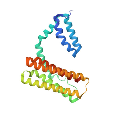
Crystal structure of human programmed cell death 10 complexed with inositol-(1,3,4,5)-tetrakisphosphate: a novel adaptor protein involved in human cerebral cavernous malformation.

Ding, J.Wang, X.Li, D.F.Hu, Y.Zhang, Y.Wang, D.C.

(2010) Biochem Biophys Res Commun 399: 587-592

  • DOI: https://doi.org/10.1016/j.bbrc.2010.07.119
  • Primary Citation of Related Structures:  
    3AJM

  • PubMed Abstract: 

    Programmed cell death 10 (PDCD10) is a novel adaptor protein involved in human cerebral cavernous malformation, a common vascular lesion mostly occurring in the central nervous system. By interacting with different signal proteins, PDCD10 could regulate various physiological processes in the cell. The crystal structure of human PDCD10 complexed with inositol-(1,3,4,5)-tetrakisphosphate has been determined at 2.3A resolution. The structure reveals an integrated dimer via a unique assembly that has never been observed before. Each PDCD10 monomer contains two independent domains: an N-terminal domain with a new fold involved in the tight dimer assembly and a C-terminal four-helix bundle domain that closely resembles the focal adhesion targeting domain of focal adhesion kinase. An eight-residue flexible linker connects the two domains, potentially conferring mobility onto the C-terminal domain, resulting in the conformational variability of PDCD10. A variable basic cleft on the top of the dimer interface binds to phosphatidylinositide and regulates the intracellular localization of PDCD10. Two potential sites, respectively located on the two domains, are critical for recruiting different binding partners, such as germinal center kinase III proteins and the focal adhesion protein paxillin.


  • Organizational Affiliation

    National Laboratory of Biomacromolecules, Institute of Biophysics, Chinese Academy of Sciences, Beijing 100101, People's Republic of China.


Macromolecules
Find similar proteins by:  (by identity cutoff)  |  3D Structure
Entity ID: 1
MoleculeChains Sequence LengthOrganismDetailsImage
Programmed cell death protein 10
A, B
213Homo sapiensMutation(s): 0 
Gene Names: PDCD10
Membrane Entity: Yes 
UniProt & NIH Common Fund Data Resources
Find proteins for Q9BUL8 (Homo sapiens)
Explore Q9BUL8 
Go to UniProtKB:  Q9BUL8
PHAROS:  Q9BUL8
GTEx:  ENSG00000114209 
Entity Groups  
Sequence Clusters30% Identity50% Identity70% Identity90% Identity95% Identity100% Identity
UniProt GroupQ9BUL8
Sequence Annotations
Expand
  • Reference Sequence
Small Molecules
Ligands 1 Unique
IDChains Name / Formula / InChI Key2D Diagram3D Interactions
4IP
Query on 4IP

Download Ideal Coordinates CCD File 
C [auth A]INOSITOL-(1,3,4,5)-TETRAKISPHOSPHATE
C6 H16 O18 P4
CIPFCGZLFXVXBG-CNWJWELYSA-N
Experimental Data & Validation

Experimental Data

  • Method: X-RAY DIFFRACTION
  • Resolution: 2.30 Å
  • R-Value Free: 0.264 
  • R-Value Work: 0.213 
  • R-Value Observed: 0.216 
  • Space Group: P 41 21 2
Unit Cell:
Length ( Å )Angle ( ˚ )
a = 89.83α = 90
b = 89.83β = 90
c = 114.2γ = 90
Software Package:
Software NamePurpose
ADSCdata collection
SOLVEphasing
PHENIXrefinement
MOSFLMdata reduction
SCALAdata scaling

Structure Validation

View Full Validation Report


Ligand Structure Quality Assessment 


Entry History 

Deposition Data

Revision History  (Full details and data files)

  • Version 1.0: 2010-06-30
    Type: Initial release
  • Version 1.1: 2011-07-13
    Changes: Version format compliance
  • Version 1.2: 2012-03-21
    Changes: Database references
  • Version 1.3: 2024-03-13
    Changes: Data collection, Database references, Derived calculations