3AJB

Crystal Structure of human Pex3p in complex with N-terminal Pex19p peptide


Experimental Data Snapshot

  • Method: X-RAY DIFFRACTION
  • Resolution: 2.50 Å
  • R-Value Free: 0.254 
  • R-Value Work: 0.219 
  • R-Value Observed: 0.221 

wwPDB Validation   3D Report Full Report


This is version 1.3 of the entry. See complete history


Literature

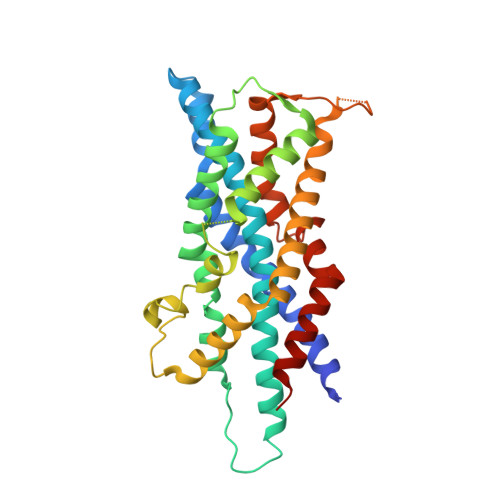
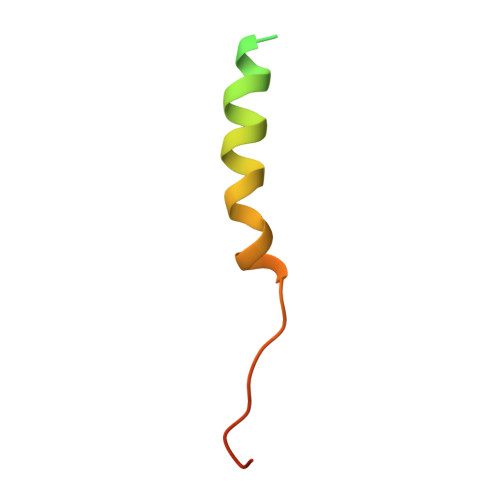
Structural basis for docking of peroxisomal membrane protein carrier Pex19p onto its receptor Pex3p

Sato, Y.Shibata, H.Nakatsu, T.Nakano, H.Kashiwayama, Y.Imanaka, T.Kato, H.

(2010) EMBO J 29: 4083-4093

  • DOI: https://doi.org/10.1038/emboj.2010.293
  • Primary Citation of Related Structures:  
    3AJB

  • PubMed Abstract: 

    Peroxisomes require peroxin (Pex) proteins for their biogenesis. The interaction between Pex3p, which resides on the peroxisomal membrane, and Pex19p, which resides in the cytosol, is crucial for peroxisome formation and the post-translational targeting of peroxisomal membrane proteins (PMPs). It is not known how Pex3p promotes the specific interaction with Pex19p for the purpose of PMP translocation. Here, we present the three-dimensional structure of the complex between a cytosolic domain of Pex3p and the binding-region peptide of Pex19p. The overall shape of Pex3p is a prolate spheroid with a novel fold, the 'twisted six-helix bundle.' The Pex19p-binding site is at an apex of the Pex3p spheroid. A 16-residue region of the Pex19p peptide forms an α-helix and makes a contact with Pex3p; this helix is disordered in the unbound state. The Pex19p peptide contains a characteristic motif, consisting of the leucine triad (Leu18, Leu21, Leu22), and Phe29, which are critical for the Pex3p binding and peroxisome biogenesis.


  • Organizational Affiliation

    Department of Structural Biology, Graduate School of Pharmaceutical Sciences, Kyoto University, Kyoto, Japan.


Macromolecules
Find similar proteins by:  (by identity cutoff)  |  3D Structure
Entity ID: 1
MoleculeChains Sequence LengthOrganismDetailsImage
Peroxisomal biogenesis factor 3330Homo sapiensMutation(s): 0 
Gene Names: hCG_18102PEX3TRG18
UniProt & NIH Common Fund Data Resources
Find proteins for P56589 (Homo sapiens)
Explore P56589 
Go to UniProtKB:  P56589
PHAROS:  P56589
GTEx:  ENSG00000034693 
Entity Groups  
Sequence Clusters30% Identity50% Identity70% Identity90% Identity95% Identity100% Identity
UniProt GroupP56589
Sequence Annotations
Expand
  • Reference Sequence
Find similar proteins by:  (by identity cutoff)  |  3D Structure
Entity ID: 2
MoleculeChains Sequence LengthOrganismDetailsImage
Peroxisomal biogenesis factor 1949Homo sapiensMutation(s): 1 
Gene Names: HK33OK/SW-cl.22PEX19PXF
UniProt & NIH Common Fund Data Resources
Find proteins for P40855 (Homo sapiens)
Explore P40855 
Go to UniProtKB:  P40855
PHAROS:  P40855
GTEx:  ENSG00000162735 
Entity Groups  
Sequence Clusters30% Identity50% Identity70% Identity90% Identity95% Identity100% Identity
UniProt GroupP40855
Sequence Annotations
Expand
  • Reference Sequence
Experimental Data & Validation

Experimental Data

  • Method: X-RAY DIFFRACTION
  • Resolution: 2.50 Å
  • R-Value Free: 0.254 
  • R-Value Work: 0.219 
  • R-Value Observed: 0.221 
  • Space Group: P 65 2 2
Unit Cell:
Length ( Å )Angle ( ˚ )
a = 147.6α = 90
b = 147.6β = 90
c = 86.202γ = 120
Software Package:
Software NamePurpose
DENZOdata reduction
SCALEPACKdata scaling
REFMACrefinement
PDB_EXTRACTdata extraction
HKL-2000data collection
HKL-2000data reduction
HKL-2000data scaling
PHENIXphasing

Structure Validation

View Full Validation Report


Entry History 

Deposition Data

Revision History  (Full details and data files)

  • Version 1.0: 2010-12-22
    Type: Initial release
  • Version 1.1: 2011-07-13
    Changes: Version format compliance
  • Version 1.2: 2017-10-11
    Changes: Refinement description
  • Version 1.3: 2024-03-13
    Changes: Data collection, Database references