3AGN

Crystal Structure of Ustilago sphaerogena Ribonuclease U2 Complexed with adenosine 3'-monophosphate


Experimental Data Snapshot

  • Method: X-RAY DIFFRACTION
  • Resolution: 0.96 Å
  • R-Value Free: 0.127 
  • R-Value Work: 0.117 
  • R-Value Observed: 0.118 

wwPDB Validation   3D Report Full Report


Ligand Structure Quality Assessment 


This is version 1.3 of the entry. See complete history


Literature

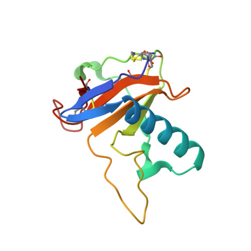
Isomerization mechanism of aspartate to isoaspartate implied by structures of Ustilago sphaerogena ribonuclease U2 complexed with adenosine 3'-monophosphate

Noguchi, S.

(2010) Acta Crystallogr D Biol Crystallogr 66: 843-849

  • DOI: https://doi.org/10.1107/S0907444910019621
  • Primary Citation of Related Structures:  
    3AGN, 3AGO

  • PubMed Abstract: 

    Aspartates in proteins are isomerized non-enzymatically to isoaspartate via succinimide in vitro and in vivo. In order to elucidate the mechanism of isoaspartate formation within the Asp45-Glu46 sequence of Ustilago sphaerogena ribonuclease U2 based on three-dimensional structure, crystal structures of ribonuclease U2 complexed with adenosine 3'-monophosphate have been solved at 0.96 and 0.99 A resolution. The crystal structures revealed that the C(gamma) atom of Asp45 is located just beside the main-chain N atom of Glu46 and that the conformation which is suitable for succinimide formation is stabilized by a hydrogen-bond network mediated by water molecules 190, 219 and 220. These water molecules are suggested to promote the formation of isoaspartate via succinimide: in the succinimide-formation reaction water 219 receives a proton from the N atom of Glu46 as a general base and waters 190 and 220 stabilize the tetrahedral intermediate, and in the succinimide-hydrolysis reaction water 219 provides a proton for the N atom of Glu46 as a general acid. The purine-base recognition scheme of ribonuclease U2 is also discussed.


  • Organizational Affiliation

    Graduate School of Pharmaceutical Sciences, The University of Tokyo, Bunkyo-ku, Tokyo, Japan. snoguchi@mol.f.u-tokyo.ac.jp


Macromolecules
Find similar proteins by:  (by identity cutoff)  |  3D Structure
Entity ID: 1
MoleculeChains Sequence LengthOrganismDetailsImage
Ribonuclease U2114Ustilago sphaerogenaMutation(s): 0 
EC: 3.1.27.4
UniProt
Find proteins for P00654 (Ustilago sphaerogena)
Explore P00654 
Go to UniProtKB:  P00654
Entity Groups  
Sequence Clusters30% Identity50% Identity70% Identity90% Identity95% Identity100% Identity
UniProt GroupP00654
Sequence Annotations
Expand
  • Reference Sequence
Small Molecules
Ligands 2 Unique
IDChains Name / Formula / InChI Key2D Diagram3D Interactions
3AM
Query on 3AM

Download Ideal Coordinates CCD File 
B [auth A][(2R,3S,4R,5R)-5-(6-aminopurin-9-yl)-4-hydroxy-2-(hydroxymethyl)oxolan-3-yl] dihydrogen phosphate
C10 H14 N5 O7 P
LNQVTSROQXJCDD-KQYNXXCUSA-N
CA
Query on CA

Download Ideal Coordinates CCD File 
C [auth A]CALCIUM ION
Ca
BHPQYMZQTOCNFJ-UHFFFAOYSA-N
Experimental Data & Validation

Experimental Data

  • Method: X-RAY DIFFRACTION
  • Resolution: 0.96 Å
  • R-Value Free: 0.127 
  • R-Value Work: 0.117 
  • R-Value Observed: 0.118 
  • Space Group: P 1 21 1
Unit Cell:
Length ( Å )Angle ( ˚ )
a = 37.88α = 90
b = 39.789β = 114.99
c = 35.884γ = 90
Software Package:
Software NamePurpose
REFMACrefinement
PDB_EXTRACTdata extraction
HKL-2000data collection
HKL-2000data reduction
SCALEPACKdata scaling
MOLREPphasing

Structure Validation

View Full Validation Report


Ligand Structure Quality Assessment 


Entry History 

Deposition Data

  • Released Date: 2010-07-07 
  • Deposition Author(s): Noguchi, S.

Revision History  (Full details and data files)

  • Version 1.0: 2010-07-07
    Type: Initial release
  • Version 1.1: 2011-07-13
    Changes: Version format compliance
  • Version 1.2: 2012-04-04
    Changes: Database references
  • Version 1.3: 2023-11-01
    Changes: Data collection, Database references, Derived calculations, Refinement description