2VF8

Crystal structure of UvrA2 from Deinococcus radiodurans


Experimental Data Snapshot

  • Method: X-RAY DIFFRACTION
  • Resolution: 3.00 Å
  • R-Value Free: 0.294 
  • R-Value Work: 0.217 
  • R-Value Observed: 0.221 

wwPDB Validation   3D Report Full Report


Ligand Structure Quality Assessment 


This is version 1.3 of the entry. See complete history


Literature

Structural and Mutational Analyses of Deinococcus Radiodurans Uvra2 Provide Insight Into DNA Binding and Damage Recognition by Uvras.

Timmins, J.Gordon, E.Caria, S.Leonard, G.Acajjaoui, S.Kuo, M.S.Monchois, V.Mcsweeney, S.

(2009) Structure 17: 547

  • DOI: https://doi.org/10.1016/j.str.2009.02.008
  • Primary Citation of Related Structures:  
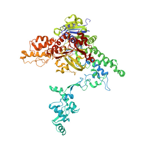
    2VF7, 2VF8

  • PubMed Abstract: 

    UvrA proteins are key actors in DNA damage repair and play an essential role in prokaryotic nucleotide excision repair (NER), a pathway that is unique in its ability to remove a broad spectrum of DNA lesions. Understanding the DNA binding and damage recognition activities of the UvrA family is a critical component for establishing the molecular basis of this process. Here we report the structure of the class II UvrA2 from Deinococcus radiodurans in two crystal forms. These structures, coupled with mutational analyses and comparison with the crystal structure of class I UvrA from Bacillus stearothermophilus, suggest a previously unsuspected role for the identified insertion domains of UvrAs in both DNA binding and damage recognition. Taken together, the available information suggests a model for how UvrA interacts with DNA and thus sheds new light on the molecular mechanisms underlying the role of UvrA in the early steps of NER.


  • Organizational Affiliation

    European Synchrotron Radiation Facility, 38043 Grenoble, France.


Macromolecules
Find similar proteins by:  (by identity cutoff)  |  3D Structure
Entity ID: 1
MoleculeChains Sequence LengthOrganismDetailsImage
EXCINUCLEASE ABC SUBUNIT A
A, B
842Deinococcus radioduransMutation(s): 1 
UniProt
Find proteins for Q9RYW8 (Deinococcus radiodurans (strain ATCC 13939 / DSM 20539 / JCM 16871 / CCUG 27074 / LMG 4051 / NBRC 15346 / NCIMB 9279 / VKM B-1422 / R1))
Explore Q9RYW8 
Go to UniProtKB:  Q9RYW8
Entity Groups  
Sequence Clusters30% Identity50% Identity70% Identity90% Identity95% Identity100% Identity
UniProt GroupQ9RYW8
Sequence Annotations
Expand
  • Reference Sequence
Small Molecules
Ligands 4 Unique
IDChains Name / Formula / InChI Key2D Diagram3D Interactions
ADP
Query on ADP

Download Ideal Coordinates CCD File 
C [auth A],
D [auth A],
J [auth B],
K [auth B]
ADENOSINE-5'-DIPHOSPHATE
C10 H15 N5 O10 P2
XTWYTFMLZFPYCI-KQYNXXCUSA-N
SO4
Query on SO4

Download Ideal Coordinates CCD File 
G [auth A]
H [auth A]
I [auth A]
N [auth B]
O [auth B]
G [auth A],
H [auth A],
I [auth A],
N [auth B],
O [auth B],
P [auth B],
Q [auth B],
R [auth B]
SULFATE ION
O4 S
QAOWNCQODCNURD-UHFFFAOYSA-L
PO4
Query on PO4

Download Ideal Coordinates CCD File 
S [auth B]PHOSPHATE ION
O4 P
NBIIXXVUZAFLBC-UHFFFAOYSA-K
ZN
Query on ZN

Download Ideal Coordinates CCD File 
E [auth A],
F [auth A],
L [auth B],
M [auth B]
ZINC ION
Zn
PTFCDOFLOPIGGS-UHFFFAOYSA-N
Experimental Data & Validation

Experimental Data

  • Method: X-RAY DIFFRACTION
  • Resolution: 3.00 Å
  • R-Value Free: 0.294 
  • R-Value Work: 0.217 
  • R-Value Observed: 0.221 
  • Space Group: C 2 2 21
Unit Cell:
Length ( Å )Angle ( ˚ )
a = 149.632α = 90
b = 171.04β = 90
c = 204.205γ = 90
Software Package:
Software NamePurpose
REFMACrefinement
MOSFLMdata reduction
SCALAdata scaling
MOLREPphasing

Structure Validation

View Full Validation Report


Ligand Structure Quality Assessment 


Entry History 

Deposition Data

Revision History  (Full details and data files)

  • Version 1.0: 2008-12-16
    Type: Initial release
  • Version 1.1: 2011-05-08
    Changes: Version format compliance
  • Version 1.2: 2011-07-13
    Changes: Version format compliance
  • Version 1.3: 2023-12-13
    Changes: Data collection, Database references, Derived calculations, Other, Refinement description