2NXW

Crystal structure of phenylpyruvate decarboxylase of Azospirillum brasilense


Experimental Data Snapshot

  • Method: X-RAY DIFFRACTION
  • Resolution: 1.50 Å
  • R-Value Free: 0.196 
  • R-Value Work: 0.175 
  • R-Value Observed: 0.175 

wwPDB Validation   3D Report Full Report


Ligand Structure Quality Assessment 


This is version 1.3 of the entry. See complete history


Literature

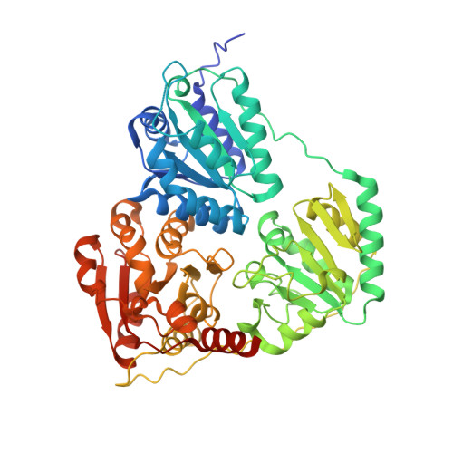
The crystal structure of phenylpyruvate decarboxylase from Azospirillum brasilense at 1.5 A resolution. Implications for its catalytic and regulatory mechanism.

Versees, W.Spaepen, S.Vanderleyden, J.Steyaert, J.

(2007) FEBS J 274: 2363-2375

  • DOI: https://doi.org/10.1111/j.1742-4658.2007.05771.x
  • Primary Citation of Related Structures:  
    2NXW

  • PubMed Abstract: 

    Phenylpyruvate decarboxylase (PPDC) of Azospirillum brasilense, involved in the biosynthesis of the plant hormone indole-3-acetic acid and the antimicrobial compound phenylacetic acid, is a thiamine diphosphate-dependent enzyme that catalyses the nonoxidative decarboxylation of indole- and phenylpyruvate. Analogous to yeast pyruvate decarboxylases, PPDC is subject to allosteric substrate activation, showing sigmoidal v versus [S] plots. The present paper reports the crystal structure of this enzyme determined at 1.5 A resolution. The subunit architecture of PPDC is characteristic for other members of the pyruvate oxidase family, with each subunit consisting of three domains with an open alpha/beta topology. An active site loop, bearing the catalytic residues His112 and His113, could not be modelled due to flexibility. The biological tetramer is best described as an asymmetric dimer of dimers. A cysteine residue that has been suggested as the site for regulatory substrate binding in yeast pyruvate decarboxylase is not conserved, requiring a different mechanism for allosteric substrate activation in PPDC. Only minor changes occur in the interactions with the cofactors, thiamine diphosphate and Mg2+, compared to pyruvate decarboxylase. A greater diversity is observed in the substrate binding pocket accounting for the difference in substrate specificity. Moreover, a catalytically important glutamate residue conserved in nearly all decarboxylases is replaced by a leucine in PPDC. The consequences of these differences in terms of the catalytic and regulatory mechanism of PPDC are discussed.


  • Organizational Affiliation

    Department of Ultrastructure, Vrije Universiteit Brussel, Brussels, Belgium. wversees@vub.ac.be


Macromolecules
Find similar proteins by:  (by identity cutoff)  |  3D Structure
Entity ID: 1
MoleculeChains Sequence LengthOrganismDetailsImage
PHENYL-3-PYRUVATE DECARBOXYLASE
A, B
565Azospirillum brasilenseMutation(s): 0 
Gene Names: ipdC
EC: 4.1.1.43
UniProt
Find proteins for P51852 (Azospirillum brasilense)
Explore P51852 
Go to UniProtKB:  P51852
Entity Groups  
Sequence Clusters30% Identity50% Identity70% Identity90% Identity95% Identity100% Identity
UniProt GroupP51852
Sequence Annotations
Expand
  • Reference Sequence
Small Molecules
Ligands 4 Unique
IDChains Name / Formula / InChI Key2D Diagram3D Interactions
TPP
Query on TPP

Download Ideal Coordinates CCD File 
E [auth A],
I [auth B]
THIAMINE DIPHOSPHATE
C12 H19 N4 O7 P2 S
AYEKOFBPNLCAJY-UHFFFAOYSA-O
GOL
Query on GOL

Download Ideal Coordinates CCD File 
F [auth A],
J [auth B],
K [auth B]
GLYCEROL
C3 H8 O3
PEDCQBHIVMGVHV-UHFFFAOYSA-N
CL
Query on CL

Download Ideal Coordinates CCD File 
D [auth A],
H [auth B]
CHLORIDE ION
Cl
VEXZGXHMUGYJMC-UHFFFAOYSA-M
MG
Query on MG

Download Ideal Coordinates CCD File 
C [auth A],
G [auth B]
MAGNESIUM ION
Mg
JLVVSXFLKOJNIY-UHFFFAOYSA-N
Experimental Data & Validation

Experimental Data

  • Method: X-RAY DIFFRACTION
  • Resolution: 1.50 Å
  • R-Value Free: 0.196 
  • R-Value Work: 0.175 
  • R-Value Observed: 0.175 
  • Space Group: C 2 2 21
Unit Cell:
Length ( Å )Angle ( ˚ )
a = 99.411α = 90
b = 179.032β = 90
c = 120.925γ = 90
Software Package:
Software NamePurpose
DENZOdata reduction
PHASERphasing
CNSrefinement
SCALEPACKdata scaling

Structure Validation

View Full Validation Report


Ligand Structure Quality Assessment 


Entry History 

Deposition Data

Revision History  (Full details and data files)

  • Version 1.0: 2007-05-29
    Type: Initial release
  • Version 1.1: 2008-05-01
    Changes: Version format compliance
  • Version 1.2: 2011-07-13
    Changes: Derived calculations, Version format compliance
  • Version 1.3: 2023-08-30
    Changes: Data collection, Database references, Derived calculations, Refinement description