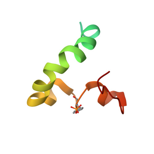
Tyrosine phosphorylation as a conformational switch: a case study of integrin Beta3 cytoplasmic tail.
Deshmukh, L., Meller, N., Alder, N., Byzova, T., Vinogradova, O.(2011) J Biol Chem 286: 40943-40953
- PubMed: 21956114
- DOI: https://doi.org/10.1074/jbc.M111.231951
- Primary Citation of Related Structures:
2LJD, 2LJE, 2LJF - PubMed Abstract:
Reversible protein phosphorylation is vital for many fundamental cellular processes. The actual impact of adding and removing phosphate group(s) is 3-fold: changes in the local/global geometry, alterations in the electrostatic potential and, as the result of both, modified protein-target interactions. Here we present a comprehensive structural investigation of the effects of phosphorylation on the conformational as well as functional states of a crucial cell surface receptor, α(IIb)β(3) integrin. We have analyzed phosphorylated (Tyr(747) and Tyr(759)) β(3) integrin cytoplasmic tail (CT) primarily by NMR, and our data demonstrate that under both aqueous and membrane-mimetic conditions, phosphorylation causes substantial conformational rearrangements. These changes originate from novel ionic interactions and revised phospholipid binding. Under aqueous conditions, the critical Tyr(747) phosphorylation prevents β(3)CT from binding to its heterodimer partner α(IIb)CT, thus likely maintaining an activated state of the receptor. This conclusion was tested in vivo and confirmed by integrin-dependent endothelial cells adhesion assay. Under membrane-mimetic conditions, phosphorylation results in a modified membrane embedding characterized by significant changes in the secondary structure pattern and the overall fold of β(3)CT. Collectively these data provide unique molecular insights into multiple regulatory roles of phosphorylation.
Organizational Affiliation:
Department of Pharmaceutical Sciences, School of Pharmacy, University of Connecticut, Storrs, Connecticut 06269, USA.