2A62

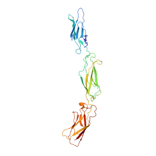
Crystal structure of mouse cadherin-8 EC1-3


Experimental Data Snapshot

  • Method: X-RAY DIFFRACTION
  • Resolution: 4.50 Å
  • R-Value Free: 0.346 
  • R-Value Work: 0.271 

wwPDB Validation   3D Report Full Report


This is version 1.3 of the entry. See complete history


Literature

Type II cadherin ectodomain structures: implications for classical cadherin specificity.

Patel, S.D.Ciatto, C.Chen, C.P.Bahna, F.Rajebhosale, M.Arkus, N.Schieren, I.Jessell, T.M.Honig, B.Price, S.R.Shapiro, L.

(2006) Cell 124: 1255-1268

  • DOI: https://doi.org/10.1016/j.cell.2005.12.046
  • Primary Citation of Related Structures:  
    1ZVN, 1ZXK, 2A4C, 2A4E, 2A62

  • PubMed Abstract: 

    Type I and II classical cadherins help to determine the adhesive specificities of animal cells. Crystal-structure determination of ectodomain regions from three type II cadherins reveals adhesive dimers formed by exchange of N-terminal beta strands between partner extracellular cadherin-1 (EC1) domains. These interfaces have two conserved tryptophan side chains that anchor each swapped strand, compared with one in type I cadherins, and include large hydrophobic regions unique to type II interfaces. The EC1 domains of type I and type II cadherins appear to encode cell adhesive specificity in vitro. Moreover, perturbation of motor neuron segregation with chimeric cadherins depends on EC1 domain identity, suggesting that this region, which includes the structurally defined adhesive interface, encodes type II cadherin functional specificity in vivo.


  • Organizational Affiliation

    Department of Biochemistry and Molecular Biophysics, Columbia University, New York, NY 10032, USA.


Macromolecules
Find similar proteins by:  (by identity cutoff)  |  3D Structure
Entity ID: 1
MoleculeChains Sequence LengthOrganismDetailsImage
Cadherin-8322Mus musculusMutation(s): 0 
Gene Names: Cdh8
UniProt
Find proteins for P97291 (Mus musculus)
Explore P97291 
Go to UniProtKB:  P97291
Entity Groups  
Sequence Clusters30% Identity50% Identity70% Identity90% Identity95% Identity100% Identity
UniProt GroupP97291
Sequence Annotations
Expand
  • Reference Sequence
Experimental Data & Validation

Experimental Data

  • Method: X-RAY DIFFRACTION
  • Resolution: 4.50 Å
  • R-Value Free: 0.346 
  • R-Value Work: 0.271 
  • Space Group: P 41 2 2
Unit Cell:
Length ( Å )Angle ( ˚ )
a = 75.818α = 90
b = 75.818β = 90
c = 233.76γ = 90
Software Package:
Software NamePurpose
REFMACrefinement
PDB_EXTRACTdata extraction
DENZOdata reduction
SCALEPACKdata scaling
PHASERphasing

Structure Validation

View Full Validation Report


Entry History 

Deposition Data

Revision History  (Full details and data files)

  • Version 1.0: 2006-04-25
    Type: Initial release
  • Version 1.1: 2008-04-30
    Changes: Version format compliance
  • Version 1.2: 2011-07-13
    Changes: Advisory, Version format compliance
  • Version 1.3: 2024-02-14
    Changes: Data collection, Database references, Derived calculations