2ZYD

Dimeric 6-phosphogluconate dehydrogenase complexed with glucose


Experimental Data Snapshot

  • Method: X-RAY DIFFRACTION
  • Resolution: 1.50 Å
  • R-Value Free: 0.191 
  • R-Value Work: 0.165 
  • R-Value Observed: 0.167 

wwPDB Validation   3D Report Full Report


Ligand Structure Quality Assessment 


This is version 1.6 of the entry. See complete history


Literature

Conformational changes associated with cofactor/substrate binding of 6-phosphogluconate dehydrogenase from Escherichia coli and Klebsiella pneumoniae: Implications for enzyme mechanism

Chen, Y.-Y.Ko, T.-P.Chen, W.-H.Lo, L.-P.Lin, C.-H.Wang, A.H.-J.

(2010) J Struct Biol 169: 25-35

  • DOI: https://doi.org/10.1016/j.jsb.2009.08.006
  • Primary Citation of Related Structures:  
    2ZYA, 2ZYD, 2ZYG, 3FWN

  • PubMed Abstract: 

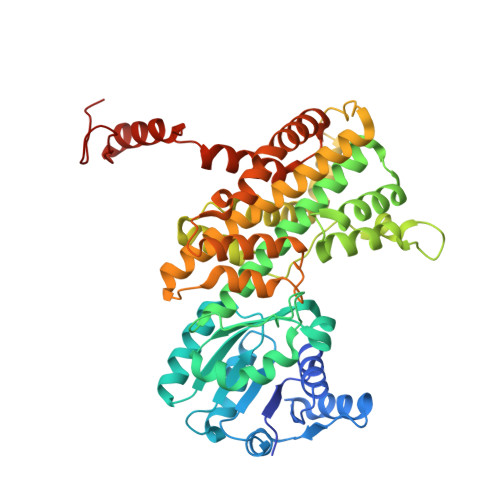
    6-Phosphogluconate dehydrogenase (6PGDH), the third enzyme of the pentose phosphate pathway, catalyzes the oxidative decarboxylation of 6-phosphogluconate, making ribulose 5-phosphate, along with the reduction of NADP(+) to NADPH and the release of CO(2). Here, we report the first apo-form crystal structure of the pathogenic Klebsiella pneumoniae 6PGDH (Kp6PGDH) and the structures of the highly homologous Escherichia coli K12 6PGDH (Ec6PGDH) complexed with substrate, substrate/NADPH and glucose at high resolution. The binding of NADPH to one subunit of the homodimeric structure triggered a 10 degrees rotation and resulting in a 7A movement of the coenzyme-binding domain. The coenzyme was thus trapped in a closed enzyme conformation, in contrast to the open conformation of the neighboring subunit. Comparison of our Ec/Kp6PGDH structures with those of other species illustrated how the domain conformation can be affected upon binding of the coenzyme, which in turn gives rise to concomitant movements of two important NADP(+)-interacting amino acids, M14 and N102. We propose that the catalysis follows an ordered binding mechanism with alternating conformational changes in the corresponding subunits, involving several related amino acid residues.


  • Organizational Affiliation

    Institute of Biological Chemistry, Academia Sinica, Taipei 115, Taiwan; Institute of Biochemical Sciences, National Taiwan University, Taipei 106, Taiwan.


Macromolecules
Find similar proteins by:  (by identity cutoff)  |  3D Structure
Entity ID: 1
MoleculeChains Sequence LengthOrganismDetailsImage
6-phosphogluconate dehydrogenase, decarboxylating
A, B
480Escherichia coli K-12Mutation(s): 1 
Gene Names: gnd
EC: 1.1.1.44
UniProt
Find proteins for P00350 (Escherichia coli (strain K12))
Explore P00350 
Go to UniProtKB:  P00350
Entity Groups  
Sequence Clusters30% Identity50% Identity70% Identity90% Identity95% Identity100% Identity
UniProt GroupP00350
Sequence Annotations
Expand
  • Reference Sequence
Small Molecules
Ligands 1 Unique
IDChains Name / Formula / InChI Key2D Diagram3D Interactions
GLO
Query on GLO

Download Ideal Coordinates CCD File 
C [auth A]D-glucose
C6 H12 O6
GZCGUPFRVQAUEE-SLPGGIOYSA-N
Experimental Data & Validation

Experimental Data

  • Method: X-RAY DIFFRACTION
  • Resolution: 1.50 Å
  • R-Value Free: 0.191 
  • R-Value Work: 0.165 
  • R-Value Observed: 0.167 
  • Space Group: P 21 21 21
Unit Cell:
Length ( Å )Angle ( ˚ )
a = 68.452α = 90
b = 118.609β = 90
c = 119.996γ = 90
Software Package:
Software NamePurpose
Blu-Icedata collection
CNSrefinement
HKL-2000data reduction
HKL-2000data scaling
CNSphasing

Structure Validation

View Full Validation Report


Ligand Structure Quality Assessment 


Entry History 

Deposition Data

Revision History  (Full details and data files)

  • Version 1.0: 2009-09-01
    Type: Initial release
  • Version 1.1: 2011-07-13
    Changes: Source and taxonomy, Version format compliance
  • Version 1.2: 2014-01-22
    Changes: Database references
  • Version 1.3: 2017-10-11
    Changes: Refinement description
  • Version 1.4: 2020-07-29
    Type: Remediation
    Reason: Carbohydrate remediation
    Changes: Database references, Derived calculations, Structure summary
  • Version 1.5: 2021-11-10
    Changes: Database references, Structure summary
  • Version 1.6: 2023-11-01
    Changes: Data collection, Refinement description