Atomic Level Description of the Domain Closure in a Dimeric Enzyme: Thermus Thermophilus 3-Isopropylmalate Dehydrogenase.
Graczer, E., Merli, A., Singh, R.K., Karuppasamy, M., Zavodszky, P., Weiss, M.S., Vas, M.(2011) Mol Biosyst 7: 1646
- PubMed: 21387033
- DOI: https://doi.org/10.1039/c0mb00346h
- Primary Citation of Related Structures:
2Y3Z, 2Y40, 2Y41, 2Y42 - PubMed Abstract:
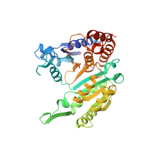
The domain closure associated with the catalytic cycle is described at an atomic level, based on pairwise comparison of the X-ray structures of homodimeric Thermus thermophilus isopropylmalate dehydrogenase (IPMDH), and on their detailed molecular graphical analysis. The structures of the apo-form without substrate and in complex with the divalent metal-ion to 1.8 Å resolution, in complexes with both Mn(2+) and 3-isopropylmalate (IPM), as well as with both Mn(2+) and NADH, were determined at resolutions ranging from 2.0 to 2.5 Å. Single crystal microspectrophotometric measurements demonstrated the presence of a functionally competent protein conformation in the crystal grown in the presence of Mn(2+) and IPM. Structural comparison of the various complexes clearly revealed the relative movement of the two domains within each subunit and allowed the identification of two hinges at the interdomain region: hinge 1 between αd and βF as well as hinge 2 between αh and βE. A detailed analysis of the atomic contacts of the conserved amino acid side-chains suggests a possible operational mechanism of these molecular hinges upon the action of the substrates. The interactions of the protein with Mn(2+) and IPM are mainly responsible for the domain closure: upon binding into the cleft of the interdomain region, the substrate IPM induces a relative movement of the secondary structural elements βE, βF, βG, αd and αh. A further special feature of the conformational change is the movement of the loop bearing the amino acid Tyr139 that precedes the interacting arm of the subunit. The tyrosyl ring rotates and moves by at least 5 Å upon IPM-binding. Thereby, new hydrophobic interactions are formed above the buried isopropyl-group of IPM. Domain closure is then completed only through subunit interactions: a loop of one subunit that is inserted into the interdomain cavity of the other subunit extends the area with the hydrophobic interactions, providing an example of the cooperativity between interdomain and intersubunit interactions.
Organizational Affiliation:
Institute of Enzymology, Biological Research Center, Hungarian Academy of Sciences, PO Box 7, H1518 Budapest, Hungary.