Consensus Structure of Pf1 Filamentous Bacteriophage from X-Ray Fibre Diffraction and Solid-State NMR.
Straus, S.K., Scott, W.R., Schwieters, C.D., Marvin, D.A.(2011) Eur Biophys J 40: 221
- PubMed: 21082179
- DOI: https://doi.org/10.1007/s00249-010-0640-9
- Primary Citation of Related Structures:
2XKM - PubMed Abstract:
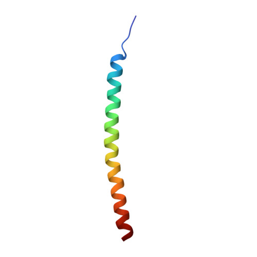
Filamentous bacteriophages (filamentous bacterial viruses or Inovirus) are simple and well-characterised macromolecular assemblies that are widely used in molecular biology and biophysics, both as paradigms for studying basic biological questions and as practical tools in areas as diverse as immunology and solid-state physics. The strains fd, M13 and f1 are virtually identical filamentous phages that infect bacteria expressing F-pili, and are sometimes grouped as the Ff phages. For historical reasons fd has often been used for structural studies, but M13 and f1 are more often used for biological experiments. Many other strains have been identified that are genetically quite distinct from Ff and yet have a similar molecular structure and life cycle. One of these, Pf1, gives the highest resolution X-ray fibre diffraction patterns known for filamentous bacteriophage. These diffraction patterns have been used in the past to derive a molecular model for the structure of the phage. Solid-state NMR experiments have been used in separate studies to derive a significantly different model of Pf1. Here we combine previously published X-ray fibre diffraction data and solid-state NMR data to give a consensus structure model for Pf1 filamentous bacteriophage, and we discuss the implications of this model for assembly of the phage at the bacterial membrane.
Organizational Affiliation:
Department of Chemistry, University of British Columbia, Vancouver, BC V6T1Z1, Canada.