2W22

Activation Mechanism of Bacterial Thermoalkalophilic Lipases


Experimental Data Snapshot

  • Method: X-RAY DIFFRACTION
  • Resolution: 2.20 Å
  • R-Value Free: 0.226 
  • R-Value Work: 0.182 
  • R-Value Observed: 0.182 

wwPDB Validation   3D Report Full Report


Ligand Structure Quality Assessment 


This is version 1.4 of the entry. See complete history


Literature

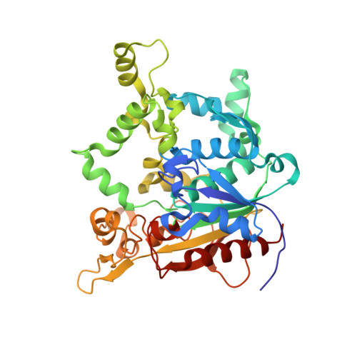
Activation of bacterial thermoalkalophilic lipases is spurred by dramatic structural rearrangements.

Carrasco-Lopez, C.Godoy, C.de Las Rivas, B.Fernandez-Lorente, G.Palomo, J.M.Guisan, J.M.Fernandez-Lafuente, R.Martinez-Ripoll, M.Hermoso, J.A.

(2009) J Biol Chem 284: 4365-4372

  • DOI: https://doi.org/10.1074/jbc.M808268200
  • Primary Citation of Related Structures:  
    2W22

  • PubMed Abstract: 

    The bacterial thermoalkalophilic lipases that hydrolyze saturated fatty acids at 60-75 degrees C and pH 8-10 are grouped as the lipase family I.5. We report here the crystal structure of the lipase from Geobacillus thermocatenulatus, the first structure of a member of the lipase family I.5 showing an open configuration. Unexpectedly, enzyme activation involves large structural rearrangements of around 70 amino acids and the concerted movement of two lids, the alpha6- and alpha7-helices, unmasking the active site. Central in the restructuring process of the lids are both the transfer of bulky hydrophobic residues out of the N-terminal end of the alpha6-helix and the incorporation of short side chain residues to the alpha6 C-terminal end. All these structural changes are stabilized by the Zn(2+)-binding domain, which is characteristic of this family of lipases. Two detergent molecules are placed in the active site, mimicking chains of the triglyceride substrate, demonstrating the position of the oxyanion hole and the three pockets that accommodate the sn-1, sn-2, and sn-3 fatty acids chains. The combination of structural and biochemical studies indicate that the lid opening is not mediated by temperature but triggered by interaction with lipid substrate.


  • Organizational Affiliation

    Grupo de Cristalografía Macromolecular y Biología Estructural, Instituto de Química-Física Rocasolano, CSIC, Serrano 119, 28006-Madrid, Spain.


Macromolecules
Find similar proteins by:  (by identity cutoff)  |  3D Structure
Entity ID: 1
MoleculeChains Sequence LengthOrganismDetailsImage
TRIACYLGLYCEROL LIPASE389Geobacillus thermocatenulatusMutation(s): 0 
EC: 3.1.1.3
UniProt
Find proteins for Q59260 (Geobacillus thermocatenulatus)
Explore Q59260 
Go to UniProtKB:  Q59260
Entity Groups  
Sequence Clusters30% Identity50% Identity70% Identity90% Identity95% Identity100% Identity
UniProt GroupQ59260
Sequence Annotations
Expand
  • Reference Sequence
Small Molecules
Ligands 4 Unique
IDChains Name / Formula / InChI Key2D Diagram3D Interactions
EGC
Query on EGC

Download Ideal Coordinates CCD File 
D [auth A],
E [auth A]
2-(2-{2-[2-(2-{2-[2-(2-{2-[4-(1,1,3,3-TETRAMETHYL-BUTYL)-PHENOXY]-ETHOXY}-ETHOXY)-ETHOXY]-ETHOXY}-ETHOXY)-ETHOXY]-ETHOX Y}-ETHOXY)-ETHANOL
C32 H58 O10
VJYAJQFKKLYARJ-UHFFFAOYSA-N
MPD
Query on MPD

Download Ideal Coordinates CCD File 
F [auth A],
G [auth A],
H [auth A]
(4S)-2-METHYL-2,4-PENTANEDIOL
C6 H14 O2
SVTBMSDMJJWYQN-YFKPBYRVSA-N
ZN
Query on ZN

Download Ideal Coordinates CCD File 
C [auth A]ZINC ION
Zn
PTFCDOFLOPIGGS-UHFFFAOYSA-N
CA
Query on CA

Download Ideal Coordinates CCD File 
B [auth A]CALCIUM ION
Ca
BHPQYMZQTOCNFJ-UHFFFAOYSA-N
Experimental Data & Validation

Experimental Data

  • Method: X-RAY DIFFRACTION
  • Resolution: 2.20 Å
  • R-Value Free: 0.226 
  • R-Value Work: 0.182 
  • R-Value Observed: 0.182 
  • Space Group: I 2 2 2
Unit Cell:
Length ( Å )Angle ( ˚ )
a = 73.07α = 90
b = 128.08β = 90
c = 127.49γ = 90
Software Package:
Software NamePurpose
CNSrefinement
MOSFLMdata reduction
SCALAdata scaling
MOLREPphasing

Structure Validation

View Full Validation Report


Ligand Structure Quality Assessment 


Entry History 

Revision History  (Full details and data files)

  • Version 1.0: 2008-12-16
    Type: Initial release
  • Version 1.1: 2011-05-08
    Changes: Version format compliance
  • Version 1.2: 2011-07-13
    Changes: Version format compliance
  • Version 1.3: 2018-02-28
    Changes: Database references, Source and taxonomy
  • Version 1.4: 2023-12-13
    Changes: Data collection, Database references, Derived calculations, Other, Refinement description, Structure summary