2QIV

Structural basis for the acyl chain selectivity and mechanism of UDP-N-acetylglucosamine acyltransferase


Experimental Data Snapshot

  • Method: X-RAY DIFFRACTION
  • Resolution: 1.85 Å
  • R-Value Free: 0.221 
  • R-Value Work: 0.191 
  • R-Value Observed: 0.193 

wwPDB Validation   3D Report Full Report


Ligand Structure Quality Assessment 


This is version 1.3 of the entry. See complete history


Literature

Structural basis for the acyl chain selectivity and mechanism of UDP-N-acetylglucosamine acyltransferase

Williams, A.H.Raetz, C.R.H.

(2007) Proc Natl Acad Sci U S A 104: 13543-13550

  • DOI: https://doi.org/10.1073/pnas.0705833104
  • Primary Citation of Related Structures:  
    2QIA, 2QIV

  • PubMed Abstract: 

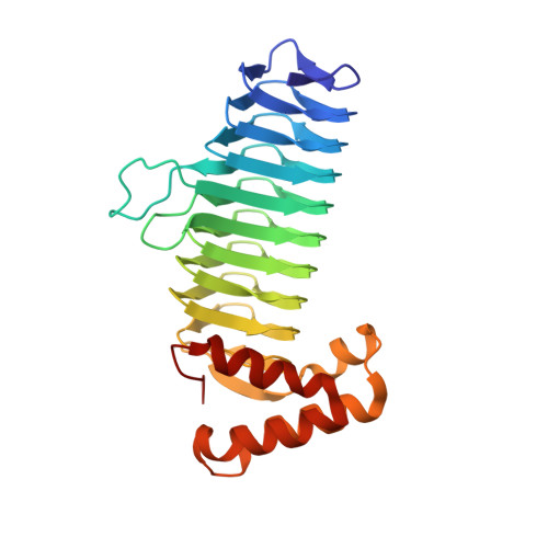
    UDP-N-acetylglucosamine (UDP-GlcNAc) acyltransferase (LpxA) catalyzes the first step of lipid A biosynthesis, the reversible transfer of the R-3-hydroxyacyl chain from R-3-hydroxyacyl acyl carrier protein to the glucosamine 3-OH group of UDP-GlcNAc. Escherichia coli LpxA is highly selective for R-3-hydroxymyristate. The crystal structure of the E. coli LpxA homotrimer, determined previously in the absence of lipid substrates or products, revealed that LpxA contains an unusual, left-handed parallel beta-helix fold. We have now solved the crystal structures of E. coli LpxA with the bound product UDP-3-O-(R-3-hydroxymyristoyl)-GlcNAc at a resolution of 1.74 A and with bound UDP-3-O-(R-3-hydroxydecanoyl)-GlcNAc at 1.85 A. The structures of these complexes are consistent with the catalytic mechanism deduced by mutagenesis and with a recent 3.0-A structure of LpxA with bound UDP-GlcNAc. Our structures show how LpxA selects for 14-carbon R-3-hydroxyacyl chains and reveal two modes of UDP binding.


  • Organizational Affiliation

    Department of Biochemistry, Duke University Medical Center, Box 3711 DUMC, Durham, NC 27710, USA.


Macromolecules
Find similar proteins by:  (by identity cutoff)  |  3D Structure
Entity ID: 1
MoleculeChains Sequence LengthOrganismDetailsImage
UDP-N-acetylglucosamine acyltransferaseA [auth X]262Escherichia coli K-12Mutation(s): 0 
Gene Names: lpxa
EC: 2.3.1.129
UniProt
Find proteins for P0A722 (Escherichia coli (strain K12))
Explore P0A722 
Go to UniProtKB:  P0A722
Entity Groups  
Sequence Clusters30% Identity50% Identity70% Identity90% Identity95% Identity100% Identity
UniProt GroupP0A722
Sequence Annotations
Expand
  • Reference Sequence
Small Molecules
Ligands 1 Unique
IDChains Name / Formula / InChI Key2D Diagram3D Interactions
U21
Query on U21

Download Ideal Coordinates CCD File 
B [auth X]uridine-5'-diphosphate-3-O-(R-3-hydroxydecanoyl)-N-acetyl-D-glucosamine
C27 H45 N3 O19 P2
MQPZMQHQQMJGDE-MCDYFTFASA-N
Experimental Data & Validation

Experimental Data

  • Method: X-RAY DIFFRACTION
  • Resolution: 1.85 Å
  • R-Value Free: 0.221 
  • R-Value Work: 0.191 
  • R-Value Observed: 0.193 
  • Space Group: P 21 3
Unit Cell:
Length ( Å )Angle ( ˚ )
a = 96.72α = 90
b = 96.72β = 90
c = 96.72γ = 90
Software Package:
Software NamePurpose
DENZOdata reduction
SCALEPACKdata scaling
MOLREPphasing
REFMACrefinement
PDB_EXTRACTdata extraction
HKL-2000data collection
HKL-2000data reduction

Structure Validation

View Full Validation Report


Ligand Structure Quality Assessment 


Entry History 

Deposition Data

Revision History  (Full details and data files)

  • Version 1.0: 2007-10-02
    Type: Initial release
  • Version 1.1: 2011-07-13
    Changes: Version format compliance
  • Version 1.2: 2017-10-18
    Changes: Refinement description
  • Version 1.3: 2024-02-21
    Changes: Data collection, Database references, Derived calculations, Structure summary