2Q8Y

Structural insight into the enzymatic mechanism of the phophothreonine lyase


Experimental Data Snapshot

  • Method: X-RAY DIFFRACTION
  • Resolution: 2.00 Å
  • R-Value Free: 0.217 
  • R-Value Work: 0.181 
  • R-Value Observed: 0.185 

wwPDB Validation   3D Report Full Report


This is version 1.4 of the entry. See complete history


Literature

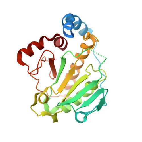
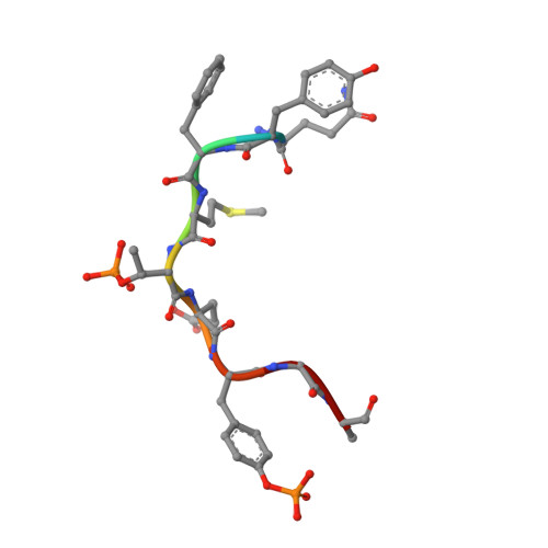
Structural insights into the enzymatic mechanism of the pathogenic MAPK phosphothreonine lyase

Zhu, Y.Li, H.Long, C.Hu, L.Xu, H.Liu, L.Chen, S.Wang, D.C.Shao, F.

(2007) Mol Cell 28: 899-913

  • DOI: https://doi.org/10.1016/j.molcel.2007.11.011
  • Primary Citation of Related Structures:  
    2P1W, 2Q8Y

  • PubMed Abstract: 

    The OspF family of phosphothreonine lyase, including SpvC from Salmonella, irreversibly inactivates the dual-phosphorylated host MAPKs (pT-X-pY) through beta elimination. We determined crystal structures of SpvC and its complex with a phosphopeptide substrate. SpvC adopts a unique fold of alpha/beta type. The disordered N terminus harbors a canonical D motif for MAPK substrate docking. The enzyme-substrate complex structure indicates that recognition of the phosphotyrosine followed by insertion of the threonine phosphate into an arginine pocket places the phosphothreonine into the enzyme active site. This requires the conformational flexibility of pT-X-pY, which suggests that p38 (pT-G-pY) is likely the preferred physiological substrate. Structure-based biochemical and enzymatic analysis allows us to propose a general acid/base mechanism for beta elimination reaction catalyzed by the phosphothreonine lyase. The mechanism described here provides a structural understanding of MAPK inactivation by a family of pathogenic effectors conserved in plant and animal systems and may also open a new route for biological catalysis.


  • Organizational Affiliation

    National Institute of Biological Sciences, Beijing, 102206, China.


Macromolecules
Find similar proteins by:  (by identity cutoff)  |  3D Structure
Entity ID: 1
MoleculeChains Sequence LengthOrganismDetailsImage
27.5 kDa virulence protein241Salmonella enterica subsp. enterica serovar EnteritidisMutation(s): 1 
Gene Names: mkaDspvCvsdD
EC: 4.2
UniProt
Find proteins for P0A2N1 (Salmonella enteritidis)
Explore P0A2N1 
Go to UniProtKB:  P0A2N1
Entity Groups  
Sequence Clusters30% Identity50% Identity70% Identity90% Identity95% Identity100% Identity
UniProt GroupP0A2N1
Sequence Annotations
Expand
  • Reference Sequence

Find similar proteins by:  Sequence   |   3D Structure  

Entity ID: 2
MoleculeChains Sequence LengthOrganismDetailsImage
Mitogen-activated protein kinase 79Homo sapiensMutation(s): 2 
EC: 2.7.11.24
UniProt & NIH Common Fund Data Resources
Find proteins for Q13164 (Homo sapiens)
Explore Q13164 
Go to UniProtKB:  Q13164
PHAROS:  Q13164
GTEx:  ENSG00000166484 
Entity Groups  
UniProt GroupQ13164
Sequence Annotations
Expand
  • Reference Sequence
Small Molecules
Modified Residues  2 Unique
IDChains TypeFormula2D DiagramParent
PTR
Query on PTR
B
L-PEPTIDE LINKINGC9 H12 N O6 PTYR
TPO
Query on TPO
B
L-PEPTIDE LINKINGC4 H10 N O6 PTHR
Experimental Data & Validation

Experimental Data

  • Method: X-RAY DIFFRACTION
  • Resolution: 2.00 Å
  • R-Value Free: 0.217 
  • R-Value Work: 0.181 
  • R-Value Observed: 0.185 
  • Space Group: P 21 21 21
Unit Cell:
Length ( Å )Angle ( ˚ )
a = 37.367α = 90
b = 71.842β = 90
c = 96.06γ = 90
Software Package:
Software NamePurpose
REFMACrefinement
CrystalCleardata collection
MOSFLMdata reduction
SCALAdata scaling
PHASESphasing

Structure Validation

View Full Validation Report


Entry History 

Deposition Data

Revision History  (Full details and data files)

  • Version 1.0: 2007-12-11
    Type: Initial release
  • Version 1.1: 2011-07-13
    Changes: Derived calculations, Version format compliance
  • Version 1.2: 2021-10-20
    Changes: Database references, Derived calculations, Source and taxonomy
  • Version 1.3: 2023-08-30
    Changes: Data collection, Refinement description
  • Version 1.4: 2023-11-15
    Changes: Data collection