


The crystal structure of an aminoglycoside 6-adenyltransferase from Bacillus subtilis
Tyagi, R., Eswaramoorthy, S., Burley, S.K., Swaminathan, S.To be published.
Experimental Data Snapshot
wwPDB Validation 3D Report Full Report

Entity ID: 1 | |||||
|---|---|---|---|---|---|
| Molecule | Chains | Sequence Length | Organism | Details | Image |
| Aminoglycoside 6-adenylyltransferase | 294 | Bacillus subtilis | Mutation(s): 12 Gene Names: aadK, BSU26790 EC: 2.7.7 | 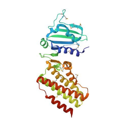 | |
UniProt | |||||
Find proteins for P17585 (Bacillus subtilis (strain 168)) Explore P17585 Go to UniProtKB: P17585 | |||||
Entity Groups | |||||
| Sequence Clusters | 30% Identity50% Identity70% Identity90% Identity95% Identity100% Identity | ||||
| UniProt Group | P17585 | ||||
Sequence AnnotationsExpand | |||||
| |||||
| Modified Residues 1 Unique | |||||
|---|---|---|---|---|---|
| ID | Chains | Type | Formula | 2D Diagram | Parent |
| MSE Query on MSE | A | L-PEPTIDE LINKING | C5 H11 N O2 Se |  | MET |
| Length ( Å ) | Angle ( ˚ ) |
|---|---|
| a = 106.562 | α = 90 |
| b = 106.56 | β = 90 |
| c = 75.601 | γ = 90 |
| Software Name | Purpose |
|---|---|
| CNS | refinement |
| CBASS | data collection |
| HKL-2000 | data reduction |
| HKL-2000 | data scaling |
| SHELX | phasing |
| SHARP | phasing |
RCSB PDB (citation) is hosted by
RCSB PDB is a member of the

