2NZH

Crystal structure of a secretion chaperone CsaA from Bacillus subtilis in the space group P 4 21 2


Experimental Data Snapshot

  • Method: X-RAY DIFFRACTION
  • Resolution: 1.90 Å
  • R-Value Free: 0.245 
  • R-Value Work: 0.200 
  • R-Value Observed: 0.202 

wwPDB Validation   3D Report Full Report


This is version 1.3 of the entry. See complete history


Literature

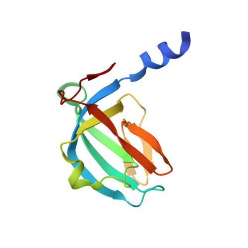
Crystallographic analysis of Bacillus subtilis CsaA.

Shapova, Y.A.Paetzel, M.

(2007) Acta Crystallogr D Biol Crystallogr 63: 478-485

  • DOI: https://doi.org/10.1107/S0907444907005045
  • Primary Citation of Related Structures:  
    2NZH, 2NZO

  • PubMed Abstract: 

    Bacillus subtilis CsaA (BsCsaA) has been proposed to act as a protein-secretion chaperone in the Sec-dependent translocation pathway, possibly compensating for the lack of SecB in the Gram-positive eubacterium Bacillus subtilis. This paper presents the cloning, purification, crystallization and structures of BsCsaA in two space groups (P42(1)2 and P3(2)21) solved and refined to resolutions of 1.9 and 2.0 A, respectively. These structures complement the previously available crystal structure of CsaA from the Gram-negative eubacterium Thermus thermophilus (TtCsaA) and provide a direct structural basis for the interpretation of previously available biochemical data on BsCsaA. The sequence and structure of the proposed substrate-binding pocket are analyzed and discussed. A comparison with the TtCsaA structure reveals a different pattern of electrostatic potential in the vicinity of the binding site, which overlaps with a region of high sequence variability. In addition, the dimerization interface of this homodimeric protein is analyzed and discussed.


  • Organizational Affiliation

    Department of Molecular Biology and Biochemistry, Simon Fraser University, South Science Building, 8888 University Drive, Burnaby, British Columbia V5A 1S6, Canada.


Macromolecules
Find similar proteins by:  (by identity cutoff)  |  3D Structure
Entity ID: 1
MoleculeChains Sequence LengthOrganismDetailsImage
Protein csaA
A, B
113Bacillus subtilisMutation(s): 0 
Gene Names: csaABSU19040
UniProt
Find proteins for P37584 (Bacillus subtilis (strain 168))
Explore P37584 
Go to UniProtKB:  P37584
Entity Groups  
Sequence Clusters30% Identity50% Identity70% Identity90% Identity95% Identity100% Identity
UniProt GroupP37584
Sequence Annotations
Expand
  • Reference Sequence
Small Molecules
Ligands 1 Unique
IDChains Name / Formula / InChI Key2D Diagram3D Interactions
GOL
Query on GOL

Download Ideal Coordinates CCD File 
C [auth A]GLYCEROL
C3 H8 O3
PEDCQBHIVMGVHV-UHFFFAOYSA-N
Experimental Data & Validation

Experimental Data

  • Method: X-RAY DIFFRACTION
  • Resolution: 1.90 Å
  • R-Value Free: 0.245 
  • R-Value Work: 0.200 
  • R-Value Observed: 0.202 
  • Space Group: P 4 21 2
Unit Cell:
Length ( Å )Angle ( ˚ )
a = 109.146α = 90
b = 109.146β = 90
c = 37.396γ = 90
Software Package:
Software NamePurpose
d*TREKdata scaling
PHASERphasing
REFMACrefinement
PDB_EXTRACTdata extraction
CrystalCleardata collection
CrystalCleardata reduction
CrystalCleardata scaling

Structure Validation

View Full Validation Report


Entry History 

Deposition Data

Revision History  (Full details and data files)

  • Version 1.0: 2007-03-27
    Type: Initial release
  • Version 1.1: 2008-05-01
    Changes: Version format compliance
  • Version 1.2: 2011-07-13
    Changes: Advisory, Version format compliance
  • Version 1.3: 2023-08-30
    Changes: Data collection, Database references, Derived calculations, Refinement description