2NOV

Breakage-reunion domain of S.pneumoniae topo IV: crystal structure of a gram-positive quinolone target


Experimental Data Snapshot

  • Method: X-RAY DIFFRACTION
  • Resolution: 2.67 Å
  • R-Value Free: 0.276 
  • R-Value Work: 0.224 

wwPDB Validation   3D Report Full Report


This is version 1.4 of the entry. See complete history


Literature

Breakage-Reunion Domain of Streptococcus pneumoniae Topoisomerase IV: Crystal Structure of a Gram-Positive Quinolone Target.

Laponogov, I.Veselkov, D.A.Sohi, M.K.Pan, X.S.Achari, A.Yang, C.Ferrara, J.D.Fisher, L.M.Sanderson, M.R.

(2007) PLoS One 2: e301-e301

  • DOI: https://doi.org/10.1371/journal.pone.0000301
  • Primary Citation of Related Structures:  
    2NOV

  • PubMed Abstract: 

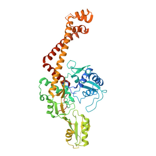
    The 2.7 A crystal structure of the 55-kDa N-terminal breakage-reunion domain of topoisomerase (topo) IV subunit A (ParC) from Streptococcus pneumoniae, the first for the quinolone targets from a gram-positive bacterium, has been solved and reveals a 'closed' dimer similar in fold to Escherichia coli DNA gyrase subunit A (GyrA), but distinct from the 'open' gate structure of Escherichia coli ParC. Unlike GyrA whose DNA binding groove is largely positively charged, the DNA binding site of ParC exhibits a distinct pattern of alternating positively and negatively charged regions coincident with the predicted positions of the grooves and phosphate backbone of DNA. Based on the ParC structure, a new induced-fit model for sequence-specific recognition of the gate (G) segment by ParC has been proposed. These features may account for the unique DNA recognition and quinolone targeting properties of pneumococcal type II topoisomerases compared to their gram-negative counterparts.


  • Organizational Affiliation

    Randall Division of Cell and Molecular Biophysics, School of Biomedical and Health Sciences, King's College London, London, United Kindgom.


Macromolecules
Find similar proteins by:  (by identity cutoff)  |  3D Structure
Entity ID: 1
MoleculeChains Sequence LengthOrganismDetailsImage
DNA topoisomerase 4 subunit A
A, B, C, D
496Streptococcus pneumoniaeMutation(s): 0 
Gene Names: parC
EC: 5.99.1
UniProt
Find proteins for Q3HYJ3 (Streptococcus pneumoniae)
Explore Q3HYJ3 
Go to UniProtKB:  Q3HYJ3
Entity Groups  
Sequence Clusters30% Identity50% Identity70% Identity90% Identity95% Identity100% Identity
UniProt GroupQ3HYJ3
Sequence Annotations
Expand
  • Reference Sequence
Experimental Data & Validation

Experimental Data

  • Method: X-RAY DIFFRACTION
  • Resolution: 2.67 Å
  • R-Value Free: 0.276 
  • R-Value Work: 0.224 
  • Space Group: I 2 2 2
Unit Cell:
Length ( Å )Angle ( ˚ )
a = 136.92α = 90
b = 137.85β = 90
c = 326.02γ = 90
Software Package:
Software NamePurpose
CNSrefinement
PDB_EXTRACTdata extraction
MAR345data collection
XDSdata reduction
XSCALEdata scaling
CNSphasing

Structure Validation

View Full Validation Report


Entry History 

Deposition Data

Revision History  (Full details and data files)

  • Version 1.0: 2006-11-14
    Type: Initial release
  • Version 1.1: 2008-05-01
    Changes: Version format compliance
  • Version 1.2: 2011-07-13
    Changes: Version format compliance
  • Version 1.3: 2017-10-18
    Changes: Refinement description
  • Version 1.4: 2023-08-30
    Changes: Data collection, Database references, Refinement description