2NO2

Crystal structure of the DLLRKN-containing coiled-coil domain of Huntingtin-interacting protein 1


Experimental Data Snapshot

  • Method: X-RAY DIFFRACTION
  • Resolution: 2.80 Å
  • R-Value Free: 0.313 
  • R-Value Work: 0.286 
  • R-Value Observed: 0.429 

wwPDB Validation   3D Report Full Report


This is version 1.2 of the entry. See complete history


Literature

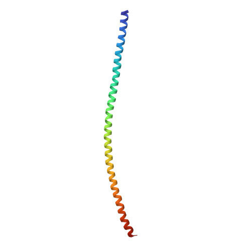
Crystal structure at 2.8 A of the DLLRKN-containing coiled-coil domain of huntingtin-interacting protein 1 (HIP1) reveals a surface suitable for clathrin light chain binding

Ybe, J.A.Mishra, S.Helms, S.Nix, J.

(2007) J Mol Biol 367: 8-15

  • DOI: https://doi.org/10.1016/j.jmb.2006.12.052
  • Primary Citation of Related Structures:  
    2NO2

  • PubMed Abstract: 

    Huntingtin interacting protein 1 (HIP1) is a member of a family of proteins whose interaction with Huntingtin is critical to prevent cells from initiating apoptosis. HIP1, and related protein HIP12/1R, can also bind to clathrin and membrane phospholipids, and HIP12/1R links the CCV to the actin cytoskeleton. HIP1 and HIP12/1R interact with the clathrin light chain EED regulatory site and stimulate clathrin lattice assembly. Here, we report the X-ray structure of the coiled-coil domain of HIP1 (residues 482-586) that includes residues crucial for binding clathrin light chain. The dimeric HIP1 crystal structure is partially splayed open. The comparison of the HIP1 model with coiled-coil predictions revealed the heptad repeat in the dimeric trunk (S2 path) is offset relative to the register of the heptad repeat from the N-terminal portion (S1 path) of the molecule. Furthermore, surface analysis showed there is a third hydrophobic path (S3) running parallel with S1 and S2. We present structural evidence supporting a role for the S3 path as an interaction surface for clathrin light chain. Finally, comparative analysis suggests the mode of binding between sla2p and clathrin light chain may be different in yeast.


  • Organizational Affiliation

    Department of Biology, Indiana University, Bloomington, IN 47405, USA.


Macromolecules
Find similar proteins by:  (by identity cutoff)  |  3D Structure
Entity ID: 1
MoleculeChains Sequence LengthOrganismDetailsImage
Huntingtin-interacting protein 1107Homo sapiensMutation(s): 0 
Gene Names: HIP1
UniProt & NIH Common Fund Data Resources
Find proteins for O00291 (Homo sapiens)
Explore O00291 
Go to UniProtKB:  O00291
PHAROS:  O00291
GTEx:  ENSG00000127946 
Entity Groups  
Sequence Clusters30% Identity50% Identity70% Identity90% Identity95% Identity100% Identity
UniProt GroupO00291
Sequence Annotations
Expand
  • Reference Sequence
Experimental Data & Validation

Experimental Data

  • Method: X-RAY DIFFRACTION
  • Resolution: 2.80 Å
  • R-Value Free: 0.313 
  • R-Value Work: 0.286 
  • R-Value Observed: 0.429 
  • Space Group: P 42 21 2
Unit Cell:
Length ( Å )Angle ( ˚ )
a = 54.2α = 90
b = 54.2β = 90
c = 152.2γ = 90
Software Package:
Software NamePurpose
d*TREKdata scaling
SOLVEphasing
RESOLVEphasing
CNSrefinement
PDB_EXTRACTdata extraction
CrystalCleardata collection
d*TREKdata reduction

Structure Validation

View Full Validation Report


Entry History 

Deposition Data

Revision History  (Full details and data files)

  • Version 1.0: 2007-10-30
    Type: Initial release
  • Version 1.1: 2011-07-13
    Changes: Version format compliance
  • Version 1.2: 2023-12-27
    Changes: Data collection, Database references