Hydroxyproline-induced Helical Disruption in Conantokin Rl-B Affects Subunit-selective Antagonistic Activities toward Ion Channels of N-Methyl-d-aspartate Receptors.
Kunda, S., Yuan, Y., Balsara, R.D., Zajicek, J., Castellino, F.J.(2015) J Biol Chem 290: 18156-18172
- PubMed: 26048991
- DOI: https://doi.org/10.1074/jbc.M115.650341
- Primary Citation of Related Structures:
2MYZ, 2MZK, 2MZL, 2MZM - PubMed Abstract:
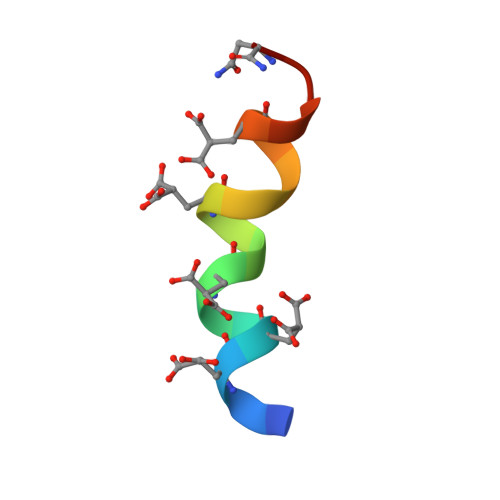
Conantokins are ~20-amino acid peptides present in predatory marine snail venoms that function as allosteric antagonists of ion channels of the N-methyl-d-aspartate receptor (NMDAR). These peptides possess a high percentage of post-/co-translationally modified amino acids, particularly γ-carboxyglutamate (Gla). Appropriately spaced Gla residues allow binding of functional divalent cations, which induces end-to-end α-helices in many conantokins. A smaller number of these peptides additionally contain 4-hydroxyproline (Hyp). Hyp should prevent adoption of the metal ion-induced full α-helix, with unknown functional consequences. To address this disparity, as well as the role of Hyp in conantokins, we have solved the high resolution three-dimensional solution structure of a Gla/Hyp-containing 18-residue conantokin, conRl-B, by high field NMR spectroscopy. We show that Hyp(10) disrupts only a small region of the α-helix of the Mn(2+)·peptide complex, which displays cation-induced α-helices on each terminus of the peptide. The function of conRl-B was examined by measuring its inhibition of NMDA/Gly-mediated current through NMDAR ion channels in mouse cortical neurons. The conRl-B displays high inhibitory selectivity for subclasses of NMDARs that contain the functionally important GluN2B subunit. Replacement of Hyp(10) with N(8)Q results in a Mg(2+)-complexed end-to-end α-helix, accompanied by attenuation of NMDAR inhibitory activity. However, replacement of Hyp(10) with Pro(10) allowed the resulting peptide to retain its inhibitory property but diminished its GluN2B specificity. Thus, these modified amino acids, in specific peptide backbones, play critical roles in their subunit-selective inhibition of NMDAR ion channels, a finding that can be employed to design NMDAR antagonists that function at ion channels of distinct NMDAR subclasses.
Organizational Affiliation:
W. M. Keck Center for Transgene Research and Department of Chemistry and Biochemistry, University of Notre Dame, Notre Dame, Indiana 46556.