Insights into the glycosaminoglycan-mediated cytotoxic mechanism of eosinophil cationic protein revealed by NMR.
Garcia-Mayoral, M.F., Canales, A., Diaz, D., Lopez-Prados, J., Moussaoui, M., de Paz, J.L., Angulo, J., Nieto, P.M., Jimenez-Barbero, J., Boix, E., Bruix, M.(2013) ACS Chem Biol 8: 144-151
- PubMed: 23025322
- DOI: https://doi.org/10.1021/cb300386v
- Primary Citation of Related Structures:
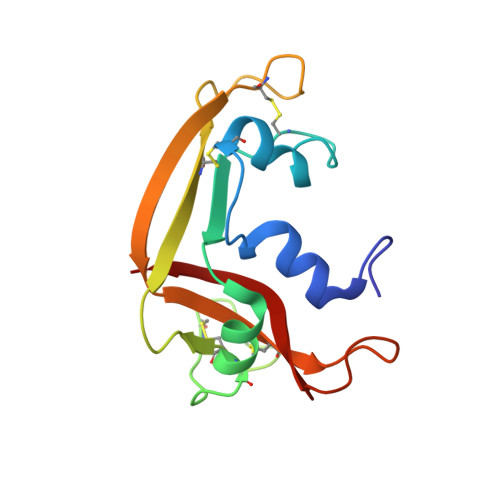
2LVZ - PubMed Abstract:
Protein-glycosaminoglycan interactions are essential in many biological processes and human diseases, yet how their recognition occurs is poorly understood. Eosinophil cationic protein (ECP) is a cytotoxic ribonuclease that interacts with glycosaminoglycans at the cell surface; this promotes the destabilization of the cellular membrane and triggers ECP's toxic activity. To understand this membrane destabilization event and the differences in the toxicity of ECP and its homologues, the high resolution solution structure of the complex between full length folded ECP and a heparin-derived trisaccharide (O-iPr-α-D-GlcNS6S-α(1-4)-L-IdoA2S-α(1-4)-D-GlcNS6S) has been solved by NMR methods and molecular dynamics simulations. The bound protein retains the tertiary structure of the free protein. The (2)S(0) conformation of the IdoA ring is preferably recognized by the protein. We have identified the precise location of the heparin binding site, dissected the specific interactions responsible for molecular recognition, and defined the structural requirements for this interaction. The structure reveals the contribution of Arg7, Gln14, and His15 in helix α1, Gln40 in strand β1, His64 in loop 4, and His128 in strand β6 in the recognition event and corroborates the previously reported participation of residues Arg34-Asn39. The participation of the catalytic triad (His15, Lys38, His128) in recognizing the heparin mimetic reveals, at atomic resolution, the mechanism of heparin's inhibition of ECP's ribonucleolytic activity. We have integrated all the available data to propose a molecular model for the membrane interaction process. The solved NMR complex provides the structural model necessary to design inhibitors to block ECP's toxicity implicated in eosinophil pathologies.
Organizational Affiliation:
Departamento de Química Física Biológica, Instituto de Química Física Rocasolano, CSIC, Madrid, Spain.